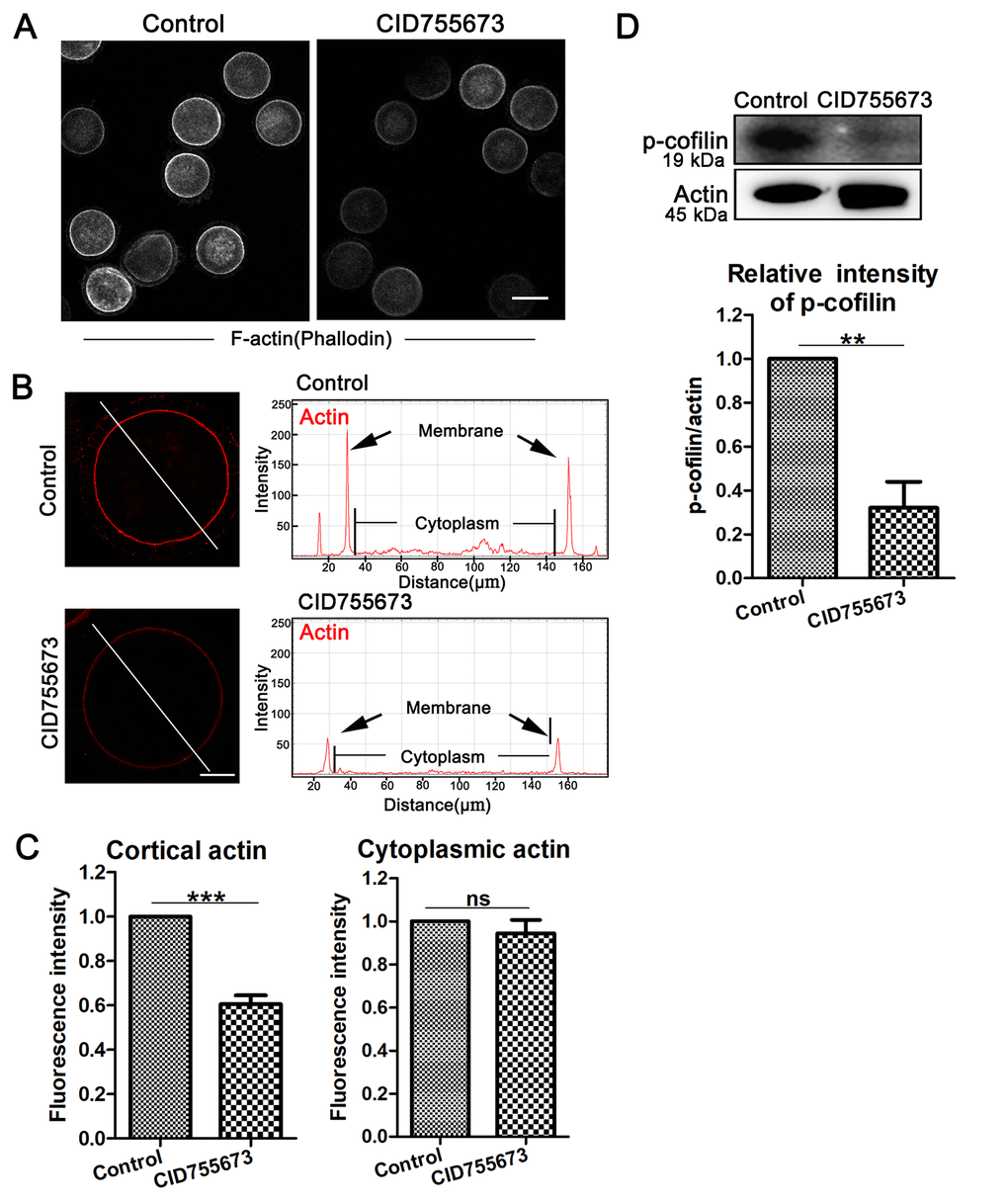
Figure 3.Effects of PKD inhibition on cortical actin assembly and p-cofilin expression during meiosis. (A) Representative images of actin distribution in the control and CID755673 treatment groups. White, actin, bar = 100 µm. (B) Fluorescence intensity profiling of actin in the left graphs. Lines in the same direction were drawn through the oocytes, and actin intensities were quantified along these lines. The black arrow in right graphs indicated actin intensity in membrane of oocyte. ZEN Blue Lite software was chosen to perform the analysis. Red, actin, bar = 30 μm. (C) Quantification immunofluorescence intensity levels of actin at the cortex and in the cytoplasm in the control and CID755673 treatment oocytes. Fluorescence intensities were analyzed using ImageJ software. (D) Protein levels of p-cofilin in control and CID755673 treatment oocytes were determined by western blotting. Data are presented as mean ± s.d. from at least three independent experiments. **, significant difference (P < 0.01). ***, significant difference (P < 0.001).