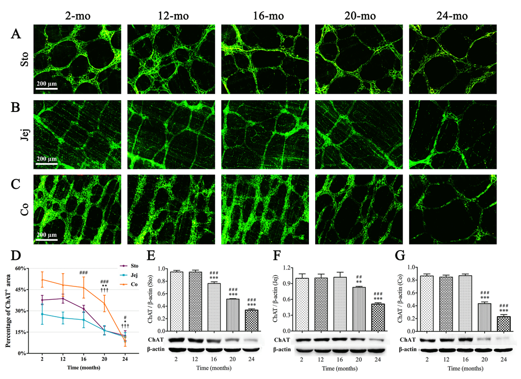
Figure 3.Decrease in ChAT(+) neurons in the MP of mouse GI tract with aging. ChAT immunoreactivity (green) was shown in ganglia and nerve fibers. ChAT-positive area per field gradually decreased from 16 mo in stomach (A), 20 mo in jejunum (B) as well as 20 mo in colon (C), respectively. Diminished immunoreactive area density (D) and the decline in expression of ChAT protein (E-G) were observed in aging mice consistent with morphological results. Densitometric analysis of protein expressions normalized to β-actin. Statistical analysis was performed using one-way analysis of variance and data were represented as mean ± SD, statistical significance is: (D) # P < 0.05, ### P < 0.001 compared with previous stomach group; * P < 0.05, ** P < 0.01 compared with previous jejunum group; ††† P < 0.001 compared with previous colon group; (E-G) ## P < 0.01, ### P < 0.001 compared with 2-mo-old group; ** P < 0.01, *** P < 0.001 compared with previous group; n=5 mice per group. Abbreviation: Sto, stomach; Jej, jejunum; Co, colon.