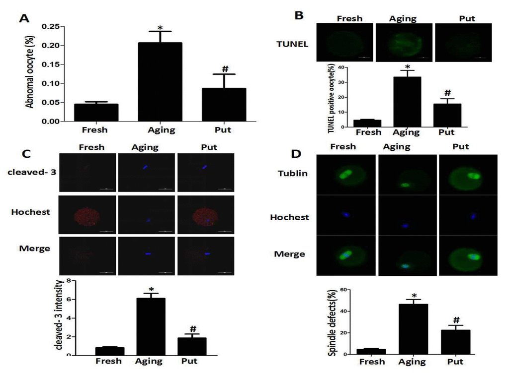
Figure 1.Putrescine decreased the apoptosis index and alleviated morphological changes during the postovulatory aging of oocytes. (A) The morphological defects of oocytes during the postovulatory aging process. The proportion of abnormal oocytes was increased in the oocytes undergoing postovulatory aging. When the MII oocytes were exposed to 0.5 mM of putrescine, the rate of morphological defects was significantly lowered when compared with the aging oocytes. (B) The level of apoptosis in the oocytes during postovulatory aging. TUNEL analysis confirmed the increased apoptosis during the postovulatory aging of oocytes. The apoptosis was significantly inhibited by the 0.5 mM of putrescine that was added to the in vitro culture medium. (C) The activated caspase 3 showed the increased level of apoptosis in the oocytes during postovulatory aging. The level of cleaved caspase 3 was significantly increased in the aging oocytes. Putrescine significantly inhibited the activation of caspase 3 in the aging oocytes. (D) The morphological observation of spindles. In the aging oocytes, the spindles became elongated, the microtubules were gradually lost from the spindle, and the aberrant chromosomal alignment was increased. The proportion of abnormal spindles was significantly decreased in the putrescine-treated group. Put, putrescine. Compared with the fresh MII oocytes, *p<0.05; compared with the aging oocytes, # p<0.05.