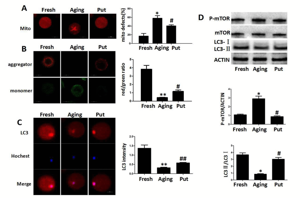
Figure 3.Functional degeneration of mitochondria in the aging oocytes. (A) The distribution of mitochondria in the cytoplasm. The even distribution of the mitochondria generally suggested a high function, while the aggregated mitochondria always showed functional degeneration. In the fresh MII oocytes, most of the mitochondria were evenly distributed in the cytoplasm. The proportion of aggregated mitochondria was significantly increased in those aging oocytes. However, this number was significantly decreased by putrescine treatment. (B) The mitochondrial membrane potential (MMP) was tested by the JC-1 assay to evaluate the mitochondrial activity. The ratio of red/green fluorescence intensity was used as the MMP index. The MMP index was significantly reduced in the aging oocytes when compared with the fresh MII oocytes. Putrescine treatment significantly enhanced the MMP of the aging oocytes. (C) The expression level of total LC3 in oocytes during postovulatory aging. The autophagy level was evaluated with total LC3 as an indicator. The autophagy in the fresh MII oocytes was at a reasonable level. The autophagy was significantly decreased in oocytes during postovulatory aging. The autophagy level was partially rescued by putrescine in the aging oocytes. (D) mTOR and LC3 transform as autophagy-related factors. The expression of mTOR was increased and the LC3-II/LC3-I ratio decreased in the aging oocytes. These effects were partially rescued by putrescine treatment in the aging oocytes. Mito, mitochondria. Put, putrescine. Compared with the fresh MII oocytes, *p<0.05, ** p<0.01; compared with the aging oocytes, # p<0.05, ## p<0.01.