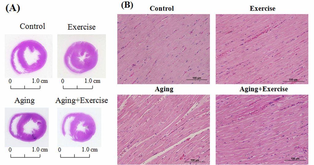
Figure 1.Hematoxylin and eosin stain (H&E stain) showing the cardiac tissue architecture. Representative histopathological analysis of cardiac sections of the left ventricles stained with hematoxylin and eosin. The hematoxylin colors basophilic structures blue-purple and the eosin colors eosinophilic structures in bright pink. The images of myocardial architecture were magnified at 400X.