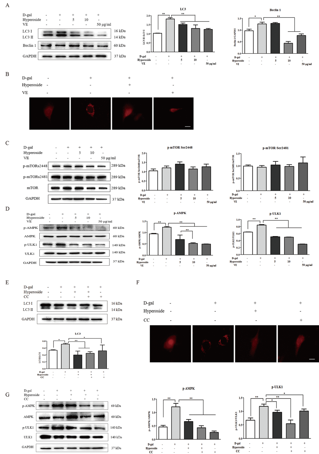
Figure 5.The effects of hyperoside and vitamin E on autophagic activity and the mTOR-independent and AMPK-ULK1 signaling pathways in vitro. (A) The NRK-52E cells were exposed to D-gal at 100 mM, with or without the treatment of hyperoside at 5 and 10 μg/ml or VE at 50 μg/ml for 24 hours, and subjected to a WB analysis for LC3 I/II and Beclin1. (B) The NRK-52E cells were infected with the RFP-LC3 Lentiviral Biosensor, exposed to D-gal at 100 mM with or without the treatment of hyperoside at 10 μg/ml or VE at 50 μg/ml for 24 hours and subjected to fluorescence microscopy. Scale bar = 5 μm. (C, D) The NRK-52E cells were exposed to D-gal at 100 mM with or without the treatment of hyperoside at 5 and 10 μg/ml or VE at 50 μg/ml for 24 hours, and subjected to a WB analysis for p-mTOR (Ser2448), p-mTOR (Ser2481) and mTOR (C1), as well as p-AMPK, AMPK, p-ULK1 and ULK1. (E) The NRK-52E cells were exposed to D-gal at 100 mM with or without the treatment of hyperoside at 10 μg/ml and CC at 10 μM for 24 hours, and subjected to a WB analysis for LC3 I/II. (F) The NRK-52E cells were infected with the RFP-LC3 Lentiviral Biosensor, exposed to D-gal at 100 mM with or without the treatment of hyperoside at 10 μg/ml and CC at 10 μM for 24 hours and subjected to fluorescence microscopy. Scale bar = 5 μm. (G) The NRK-52E cells were exposed to D-gal at 100 mM with or without the treatment of hyperoside at 10 μg/ml and CC at 10 μM for 24 hours, and subjected to a WB analysis for p-AMPK, AMPK, p-ULK1 and ULK1. The data are expressed as the mean ± SD, (n=3), *P < 0.05, **P < 0.01. Abbreviation: D-gal, D-galactose; VE, vitamin E; WB, Western blot; p-mTOR, phosphorylated mTOR; p-AMPK, phosphorylated AMPK; p-ULK1, phosphorylated ULK1; CC, compound C.