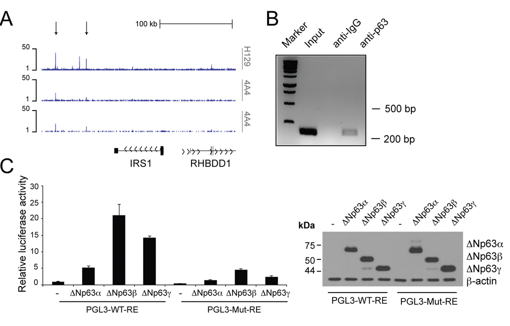
Figure 2.p63 binds to the regulatory region of theIrs1gene. (A) p63 DNA-binding profiles in the Irs1 locus, obtained in NHEKs by ChIP-sequencing (ChIP-seq) using 4A4 and H129 anti-p63 antibodies in two normal human primary keratinocyte cell lines (K1 and K2) [87]. (B) ChIP analysis of p63 occupancy at the regulatory regions of the Irs1 gene. ChIP assays were performed in Fadu HNSCC cells using H129 anti-p63 antibody and control IgGs. PCR validation was performed using primers spanning the p63-binding sites located within the genomic regions identified by ChIP-seq assays. (C) Luciferase reporter assays of Irs1 regulatory regions (left panel). The pGL3 reporter vector (30 ng) and the pRL-CMV-Renilla luciferase plasmid (5 ng) were cotransfected with the empty pcDNA-HA vector or plasmids coding ΔNp63α, ΔNp63β, and ΔNp63γ (150 ng) into the p53 null human H1299 cell line. The luciferase activities of cellular extracts were measured 24 h after transfection. Cellular lysates were also analysed by western blot (right panel). Data are presented as mean ± SD and are representative of three independent experiments.