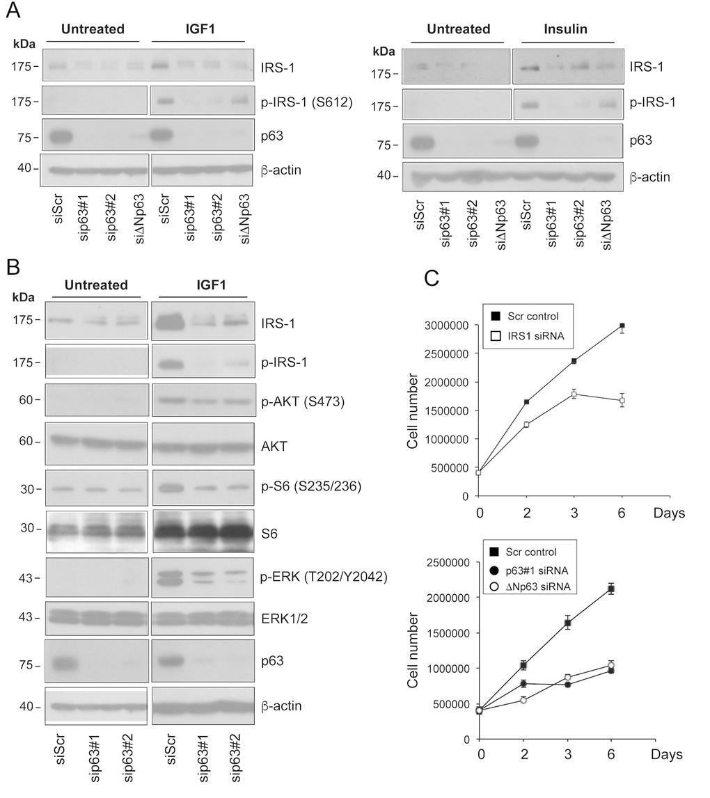
Figure 3.Depletion of p63 reduces the responsiveness of HNSCC cells to ligand stimulation. (A) Fadu cells were transfected with siScr or different p63 (sip63#1, sip63#2, siΔNp63) siRNAs. Forty-eight h after transfection, cells were serum starved for 4 h, and then stimulated with 5 nM IGF1 (upper panel) or 500 ng/ml insulin (lower panel) for 10 min. Protein amounts of p63, IRS1 and p-IRS1 were detected by western blot analysis. β-actin served as loading control. Blots are representative of three individual experiments. (B) Fadu cells were transfected with siScr, sip63#1 and sip63#2, serum starved for 4 h and then stimulated with 5 nM IGF1 for 10 min. Cellular extracts were analysed with the following antibodies: anti-IRS1, anti-p-IRS1, anti-p-AKT, anti-AKT, anti-p-S6 Ribosomal Protein, anti-S6, anti-p44/42 MAPK (p-ERK1/2), anti-ERK1/2, p63 and β-actin as loading control. Blots are representative of three individual experiments. (C) Fadu cells were transfected with siScr or siIRS1 (upper panel) and with sip63#1, ΔNp63, or siScr (lower panel). Forty-eight h after transfection, cells were seeded in 6-cm plates at 500,000/plate and growth was followed until day 6.