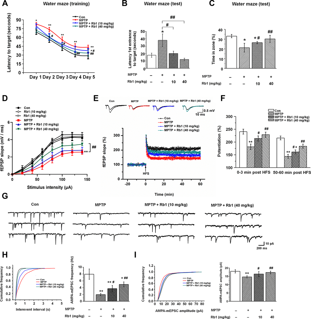
Figure 1.Rb1 prevents cognitive impairment and dysfunctional glutamatergic transmission in the MPTP mouse model of PD. (A–C) Morris water maze tests were conducted after treatment with MPTP or different doses of Rb1. Mice were analyzed for (A) the escape latency during a 5-day training course. In the probe tests, mice were analyzed (B) for the escape latency, and (C) the time spent in the target zone. n = 12 per group. (D) Input–output relations generated by stimulating the SCs and recording in CA1 stratum radiatum. n = 6–10. (E) The effect of Rb1 on the LTP at the SC-CA1 synapses was recorded in MPTP-treated mice. The middle image shows representative traces of fEPSP recordings of responses before and 50 min after high-frequency stimulation (HFS; arrow). (F) Quantitative analysis of data in e. The level of fEPSP potentiation was determined at a mean of 0–3 min and 50–60 min after high-frequency stimulation. n = 5–8. (G) Representative traces of APMA receptor-mediated mEPSCs. All mEPSCs were recorded at a holding potential of −65 mV. (H) Cumulative frequency plots of the inter-event interval (left) and quantitative analysis of the frequency of APMA receptor-mediated mEPSCs (right). (I) Cumulative frequency plots of the amplitude (left) and quantitative analysis of the amplitude of APMA receptor-mediated mEPSCs (right). n = 11–15 per group. Data were obtained from the whole-cell recordings of the pyramidal neurons in the hippocampal CA3 region from the four groups of mice. Results are expressed as the mean ± SEM. **p < 0.01, *p < 0.05 vs. control group; ##p < 0.01, #p < 0.05 vs. MPTP group. Statistical significance was determined by one-way ANOVA and Bonferroni tests as post hoc comparisons.