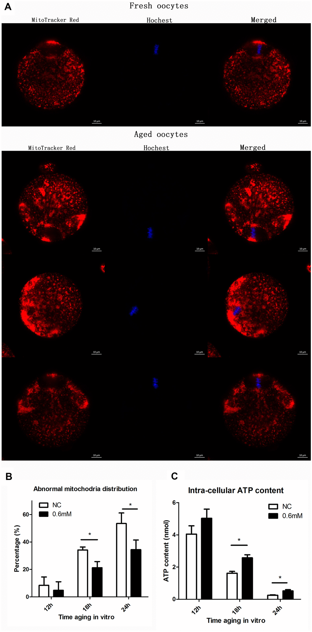
Figure 5.NAC treatment protects the function of mitochondria during post-ovulatory oocyte aging in vitro. (A) Confocal micrographs of mitochondrial distribution. Mitochondria were stained with MitoTracker-Red, and chromosomes were stained with Hoechst 33342 (blue). (B) Percentages of abnormal distribution of mitochondria in oocytes. Oocytes with normal mitochondria distribution and the those with abnormal mitochondira distribution were counted for calculating the percentage of abnormal mitochondria distribution found in oocytes in each experiment. NC, control group in which oocytes were not treated with NAC. 0.6mM, oocytes treated with 0.6mM NAC. Data are expressed as mean ± SEM of at least 3 independent experiments, and 6 superovulated mice were killed to obtain a minimum of 40 oocytes for each experiment. Star represents mean difference, 0.01< p < 0.05. (C) The adenosine triphosphate (ATP) content of mouse oocyte at different aging time points of two groups with or without NAC treatment. A Berthold Lumat LB 9501 luminometer and a commercial assay kit were used for ATP measurement. Six superovulated mice were killed to obtain a minimum of 40 oocytes to investigate the mitochondria distribution for each independent replicates, and 14 superovulated mice were killed to obtain a minimum of 100 oocytes to analyze the ATP content for each independent replicate.