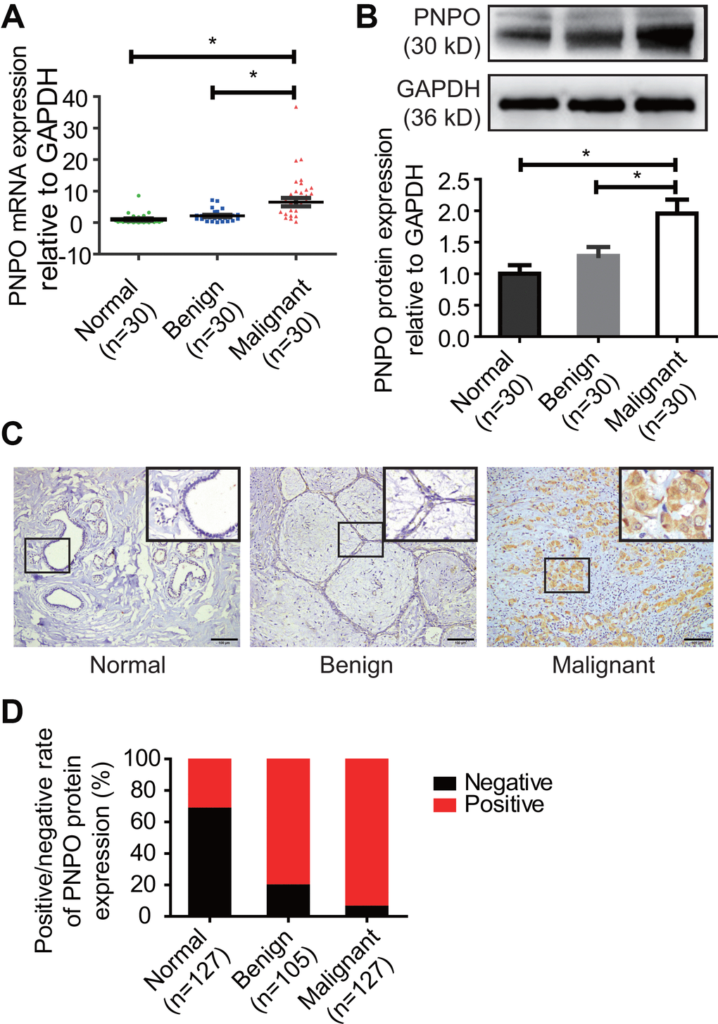
Figure 1.PNPO expression in human breast tissues. (A) PNPO mRNA expression was detected in breast tissues by qRT-PCR. (B) PNPO protein expression was detected in breast tissues by Western blot. Representative images and semi-quantitative analysis of the relative PNPO protein expression are shown in the upper and lower panel, respectively. (C) PNPO protein expression was detected in paraffin-embedded breast tissues by IHC. A picture in a frame is amplified. Brown color in a cell is considered positive staining. Original magnification, x200. Scale bar, 100 μm. (D) Quantified data of IHC. Normal, adjacent normal breast tissue; Benign, breast benign tumor (fibroadenomas); Malignant, breast malignant tumor (invasive ductal carcinoma); Negative, negative staining; positive, positive staining; n, number of cases. * P < 0.05.