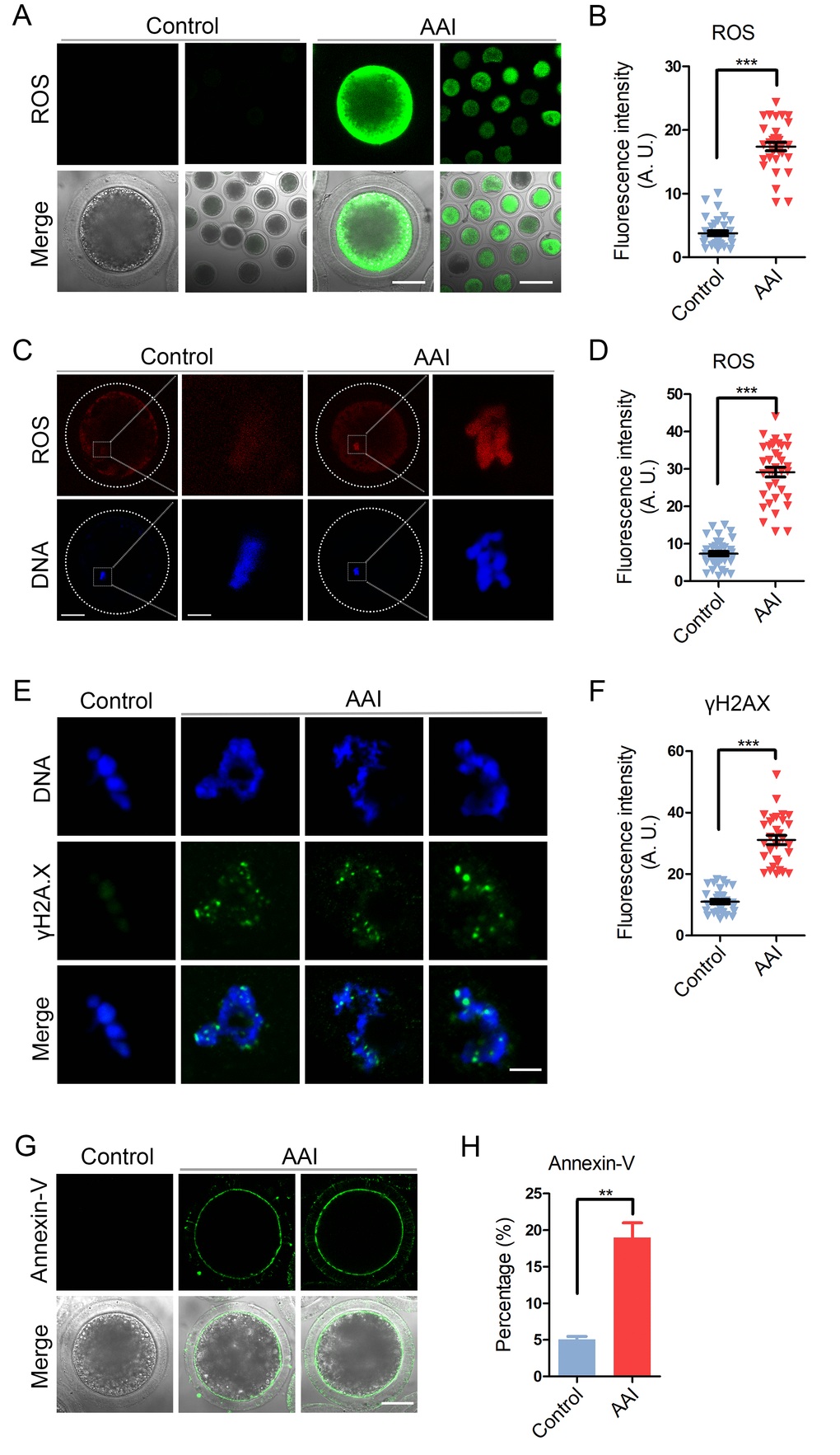
Figure 8.Effects of AAI exposure on the ROS level, DNA damage and early apoptosis in porcine oocytes. (A) Representative images of DCFH staining in control and AAI-exposed oocytes. Scale bar, 40 and 80 μm. (B) The fluorescence intensity of ROS levels was recorded in control and AAI-exposed oocytes. (C) Representative images of DHE staining in control and AAI-exposed oocytes. Scale bar, 20 and 5 μm. (D) The fluorescence intensity of ROS levels was recorded in control and AAI-exposed oocytes. (E) Representative images of DNA damage in control and AAI-exposed oocytes. Scale bar, 5 μm. (F) The fluorescence intensity of γH2AX signals was measured in control and AAI-exposed oocytes. (G) Representative images of apoptotic oocytes in control and AAI-exposed groups. Scale bar, 40 μm. (H) The rate of apoptotic oocytes was recorded in control and AAI-exposed groups. Data in (B), (D), (F) and (H) were presented as mean percentage (mean ± SEM) of at least three independent experiments. **P < 0.01, ***P < 0.001.