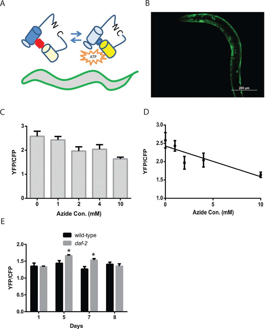
Figure 2.Reduced daf-2 insulin-like signaling increases ATP levels in C. elegans muscle. (A) Cartoon of the ATeam ratiometric reporter (Adenosine 5’-Triphosphate indicator based on Epsilon subunit for Analytical Measurement) which consists of the cyan fluorescent protein derivative mseCFP and the yellow fluorescent protein mVenus flanking the ε-subunit of from the F0F1-ATP synthase from Bacillus subtilis. The subunit from the F0F1-ATP synthase binds ATP and thereby changes in the spatial arrangement of the fluorophores which then results in alterations in the excitation of the yellow fluorophore by the cyan fluorophore via FRET. (B) Fluorescence image of a wild-type worm expressing the ATeam reporter under the control of the myo-3 promoter in the body-wall muscle. Image was captured using a GFP filter set which can visualize mVenus fluorescence. Bar: 200 µm. (C) ATeam shows a reduction in the ratio of mVenus to mseCFP fluorescence (YFP/CFP) when ATP levels are reduced by treatment with the mitochondrial inhibitor sodium azide. Worms expressing ATeam in the muscle were treated with increasing doses of sodium azide for 1 hour before being mounted and digitally imaged. N >12 for all treatments. These data also exhibit a linear decline when plotted as an X-Y graph (D). (E) The daf-2 mutants have increased muscle ATP levels on adult day 5 and day 7 as shown by the imaging of wild-type and daf-2 mutant animals expressing the ATeam reporter. Each bar represents the average ATeam YFP/CFP ratio from the muscles of daf-2 mutant and wild-type animals on the indicated days of adulthood. N >12 for all ages and genotypes. * represents p < 0.05 by t‐test.