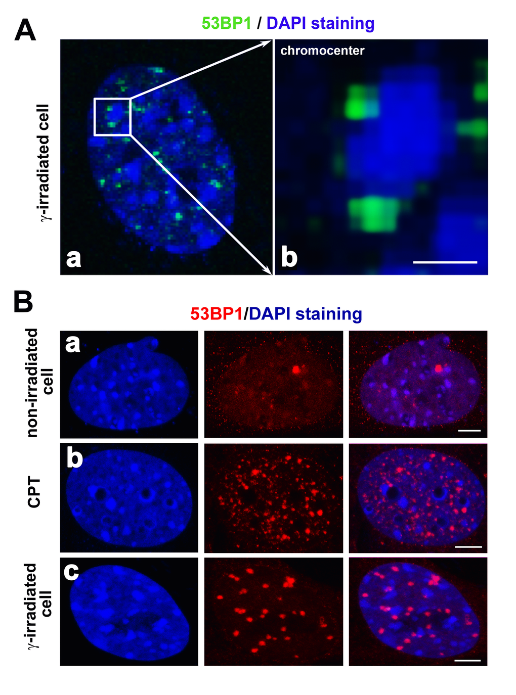
Figure 5.(A) Localization of the 53BP1 protein (green) in close proximity to chromocenters (clusters of centromeric heterochromatin; blue) is shown. Reindl et al. [68] showed that the 53BP1 protein localized in close proximity to the perichromatin region. This picture is our illustration of 53BP1 localization at the periphery of chromocenters. Here, DAPI was used for the visualization of MEF nuclei. In panel (A), chromocenters are characterized by dense DAPI staining. Panel (a) shows the DAPI-stained interphase nucleus and (b) is the magnified chromocenter (blue) decorated by 53BP1-positive foci (green). The scale bar represents 1 µm. (B) Compared to (a) non-irradiated cells, (b) tiny DNA damage foci may be induced by camptothecin (CPT) treatment. The 53BP1 protein (red) did not overlap with chromocenters in (c) γ-irradiated cells. The 53BP1 foci of CPT-treated cells were characterized by a distinct morphology compared to IRIF. A number of foci may be different in distinct cell lines and after cell exposure to distinct types of radiation or DNA damaging agents, as shown by [80] or [53], and see here. Scale bars in panels Ba-c represent 5 µm.