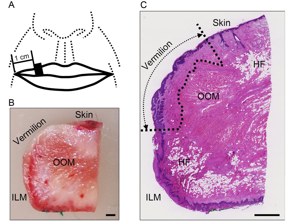
Figure 1.Areas used for investigation. (A) Specimen collection site: the black square indicates the site of specimen collection. (B) Representative image of a dissected specimen: the skin surface and intraoral labial mucosa on the oral cavity side were stained with red and green pigments, respectively. Bar=1 mm; OOM, orbicularis oris muscle; ILM, intraoral labial mucosa. (C) Representative image of hematoxylin and eosin stained section of upper lip: the boundary of the vermilion is indicated by the dotted line. Bar=1 mm; OOM, orbicularis oris muscle; HF, hypodermal fat; ILM, intraoral labial mucosa. Note: Hypodermal fat in the vermilion was absent from all specimens.