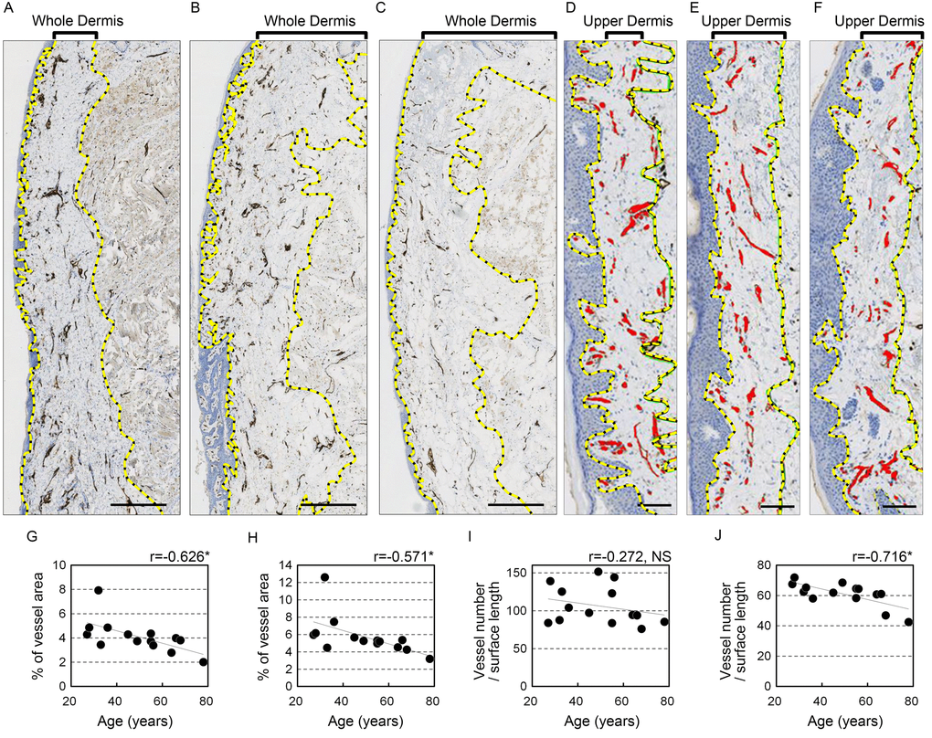
Figure 2.Blood vessels in the upper lip vermilion dermis decrease with age. Vessels are stained for anti-CD31. (A-F) Representative image staining: brown, CD31+ vessels; blue, cell nuclei. (A-C) Images of the whole vermilion dermis. Donors were 28 (A), 49 (B) and 64 (C) years old. Bar=500 μm. The yellow dotted line marks the boundary line of the whole dermis. (D-F) Images of the upper dermis of the vermilion. Donors were 27 (D), 45 (E), and 68 (F) years old. Bar=100 μm; red, blood vessels in upper dermis of the vermilion; yellow dotted line, boundary for the upper dermis. (G-K) Individual plots of vessel parameters shown according to age. (G, H) Age plotted against percentage of blood vessel area in the analyzed area. (I, J) Age plotted against the number of blood vessels per surface length (mm). (G, I) Whole vermilion dermis. (H, J) Upper dermis of the vermilion. n=14. *p<0.05 (Pearson's correlation test).