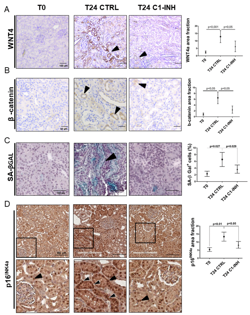
Figure 7.Wnt4/βcatenin pathway and inflammaging markers are activated in tubular cells after I/R and modulated by C1-INH treatment. (A–B) Immunoistochemical stainings showing the tubular Wnt4 and βcatenin increase after 1 day of I/R injury and the C1-INH-mediated modulation. IHC was performed on paraffin kidney sections. Arrows indicate positive tubular staining. (in the right) Graphical representation of Wnt4 and βcatenin protein expression level in the different groups. (n=5, p value as indicated, scale bar as indicated).(C) Representative SA-β Gal stained kidney tissues revealed an higher number of senescent cells after 24 h of I/R injury compared to T0. Treatment with C1-INH restored SA-β Gal at basal expression. Arrows indicate positive tubular staining on cryo tissues. (in the right) Graphical representation %SA-βGal area fraction. (n=5, p value as indicated, scale bar as indicated). (D) Representative micrographs indicating p16INK4a protein expression in different groups of swine, as indicated. Boxed areas are enlarged in the bottom of each micrographs. In the T0, p16INK4a had constitutive level and was localized in tubular nuclei (black arrow). Biopsies after 24h from reperfusion (T24 CTRL) showed increased nuclear (black arrow) and cytoplasmic staining (white dotted arrows). C1-INH restored p16INK4a at basal expression; limiting the cytoplasmic p16INK4a expression. (in the right) Graphical representation of p16INK4a area fraction in the different groups. (n=5, p value as indicated, scale bar as indicated).