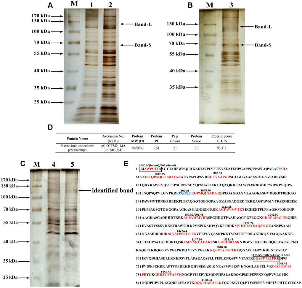
Figure 4.Purification and identification of MAP6 via lectin affinity chromatography coupled with MALDI-TOF MS. (A) SDS-PAGE analysis on the fractions from wash buffer containing 0.3 mol/L NaCl. Lanes 1 and 2 were the different fractions. Lane M, protein size standards. (B) SDS-PAGE analysis of the fractions from eluted buffer containing 0.5 mol/L NaCl. Lane 3, eluted fraction. Lane M, protein size standards. (C) SDS-PAGE analysis of purified glycoproteins and silver staining: lane M, protein size standards – the arrow shows the glycoprotein subjected to MS identification; Lanes 4 and 5, glycoproteins purified in lectin pulldown assays. (D) Profiles of identified MAP6 in Mus musculus. (E) Trypsin-digested MAP6 peptide fragments identified based on their MALDI-TOF MS spectrum. The identified peptide fragments are indicated in red, blue, and box. The numbers on the sequence are the m/z of the molecular fragment ions after collision-induced dissociation.