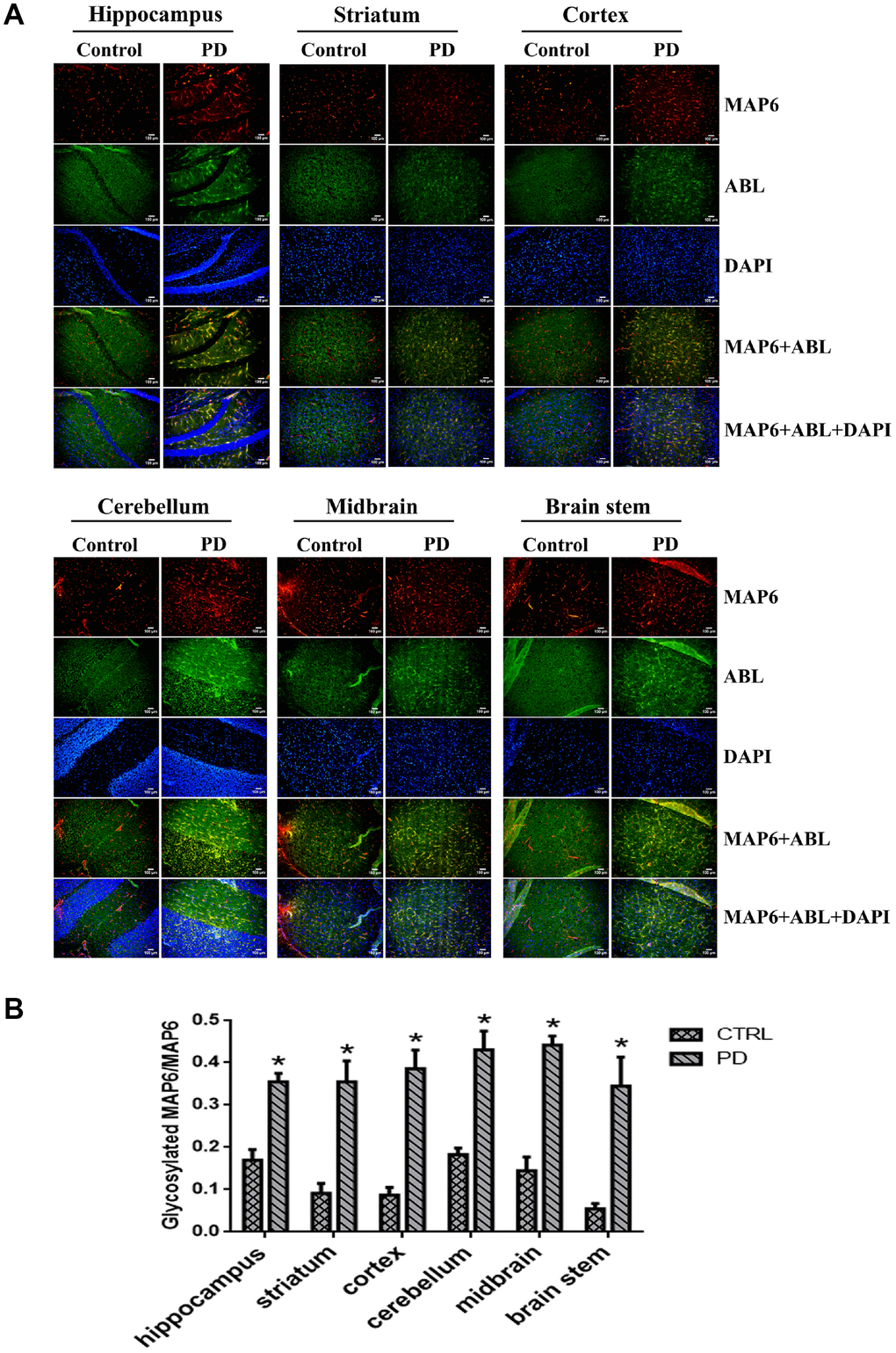
Figure 7.In situ dual immunofluorescence analysis glycosylated MAP6 in various brain areas in PD mice. (A) Representative photomicrographs of immunofluorescent labeling of glycoproteins detected by ABL (green) and MAP6 stained using anti-MAP6 monoclonal antibody (red) in the indicated areas, as well as their colocalization (yellow). Nuclei were stained using DAPI (blue). (B) Statistical comparison of the immunofluorescent signals between the MPTP-induced PD mice and control mice. Bars depict the mean±SD; *p<0.05 vs. control group.