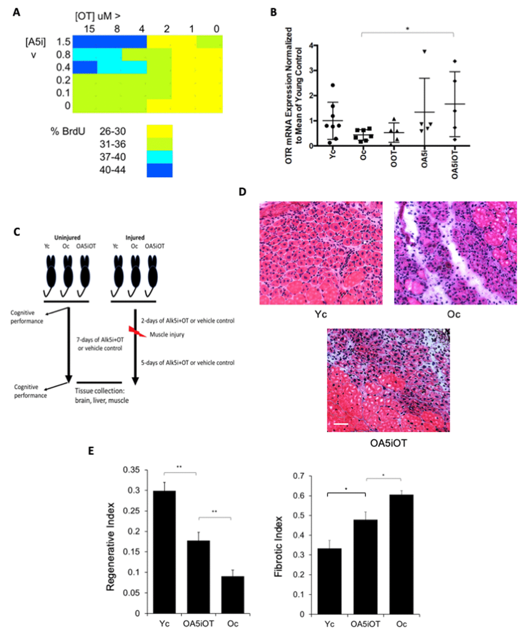
Figure 1.Effects of OT and Alk5i on myogenic proliferation, OTR expression and muscle repair in vivo. (A) Old muscle stem cells were freshly isolated from aged (23-24mo old) C57.B6 male mice and cultured in Opti-MEM with 5% old mouse serum. The indicated doses of Alk5i and OT (both micromolar) were added to 103 cells per well of 96 well plates for 24 hours. Cells were pulsed with BrdU, immunostained and counted. Shown is the percent proliferation visualized as a heat map. The effects of OT and Alk5i on proliferation are dose dependent and at some doses Alk5i+OT has a more robust effect than each molecule alone. (B) Old (23-24mo) C57.B6 mice were administered by subcutaneous injections with oxytocin (OOT), Alk5i (OA5i), a mixture of OT and Alk5i (OA5iOT) or HBSS (Oc) in vivo for 7 days, daily. Young (2-3mo) mice (Yc) were injected with HBSS in an identical manner. The expression levels of oxytocin receptor (OTR) were assayed by real-time qRT-PCR in the TA muscles of these mice and were normalized to Actin. OA5iOT as compared to Oc (*p = 0.030). N = 8 for Yc, N = 7 for Oc, N = 5 for OOT, N = 5 for OA5i, and N = 5 for OA5iOT. (C) Schematic of the experimental procedure. Old (23-24 month) C57.B6 mice were injected subcutaneously with Alk5i+OT (0.02 nmol/g/day for Alk5i, and 1 µg/g/day for OT) (OA5iOT) or control vehicle (HBSS) (Oc) for 7 days daily. The young C57.B6 mice (Yc, 3-4 month), were identically administered with HBSS for 7 days. After two days of Alk5i+OT or HBSS injections, some young and old mice underwent experimental muscle injury and were then treated with Alk5i+OT or HBSS for 5 days; while other mice were analyzed in the absence of tissue injury. Male mice were used in these studies. (D) TA muscles were injured by injections of CTX and 5 days later, muscles were snap-frozen in OCT and cryosectioned to 10 µm. H&E staining was performed where newly formed muscle fibers are smaller and with central nuclei. These nascent myofibers form efficiently in the young, but not old injured muscles. As shown in representative H&E panels, Alk5i+OT dramatically enhanced in vivo myogenesis (dense areas of new myofibers) and diminished fibrosis (white areas devoid of muscle fibers). Scale bar=50 µm. (E) The regenerative index and fibrotic indices were defined at 5 days post CTX injury, as in (Rebo J., et al 2016); Alk5i+OT improved muscle regeneration and reduced fibrosis (*p=0.02201, **p Oc & OA5iOT = 0.0029, **p Yc & OA5iOT = 0.00870). N=5 for each cohort in both regenerative and fibrotic studies.