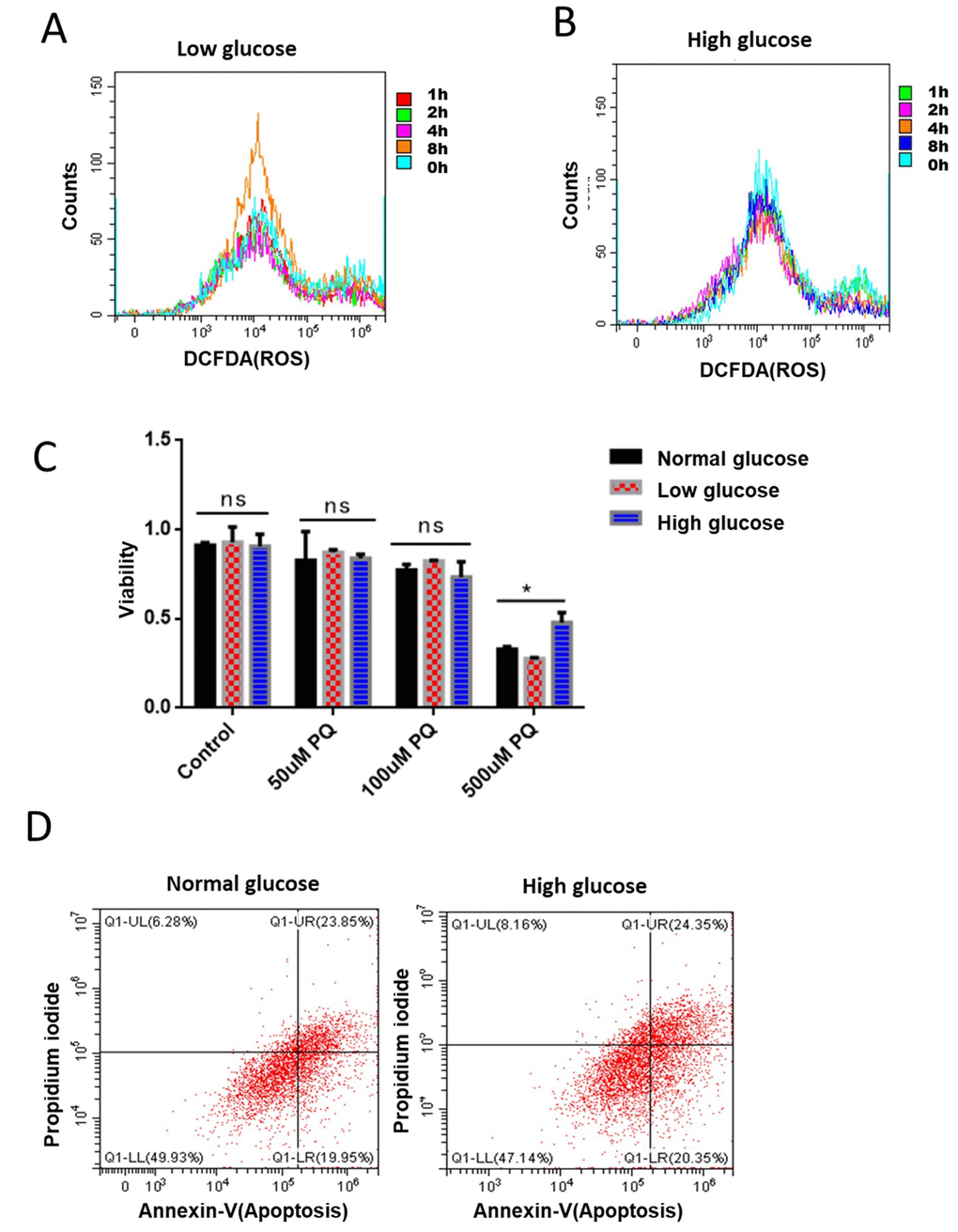
Figure 5.Glucose suppression of ROS is conserved in mammalian cells. (A) Short term rapamycin treatment increased intracellular ROS levels. Mouse embryonic fibroblasts (NIH3T3) cultured in medium supplemented with 1g/L glucose were treated with 100nM rapamycin for 1, 2, 4, and 8 hours. ROS levels were measured by staining cells with 2,7′–dichlorofluorescin diacetate (DCFDA), followed by flow cytometry analysis. (B) High glucose suppressed rapamycin-induced ROS in mouse embryonic fibroblasts. Mouse embryonic fibroblasts (NIH3T3) cultured in medium supplemented with 8g/L glucose were treated with 100nM rapamycin for 1, 2, 4, and 8 hours. ROS levels were measured by staining cells with 2,7′–dichlorofluorescin diacetate (DCFDA), followed by flow cytometry analysis. (C) High glucose suppressed ROS-induced cell death in mouse embryonic fibroblasts. NIH3T3 cells maintained in low (1g/L), normal (4g/L) and high glucose (8g/L) were treated with indicated concentrations of ROS-generator paraquat for 24 hours. Cell viability was detected by Cell Counting Kit-8 (CCK-8) assay. Means of 3 independent experiments were plotted with error bars showing the standard deviation. P value by student’s t-test: *, P<0.05. (D) Apoptosis was not significantly repressed by glucose. NIH3T3 cells maintained in normal (4g/L) and high glucose (8g/L) were treated 500μM ROS generator paraquat for 24 hours to induce cell apoptosis. Cell viability was measured by Propidium iodide (PI) staining and apoptosis were detected by Annexin V-FITC followed by flow cytometry analysis.