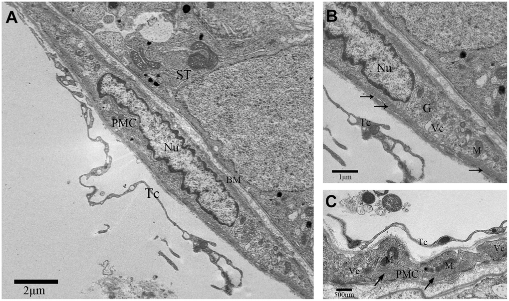
Figure 1.TEM photograph of a peritubular myoid cell in the rat testis. (A) Peritubular myoid cells are seen in the lamina propria of the seminiferous tubule. (B–C) A single peritubular myoid cell (PMC) is shown with actin filaments (indicated by the arrow), mitochondria, Golgi apparatus and secretory vesicles. Note: PMC: peritubular myoid cell; ST: seminiferous tubule; TC: telocyte; BM: basement membrane; Nu: nucleus; M: Mitochondria; G: Golgi apparatus;Vc: Vesicles. Scale Bar = A: 2μm; B: 1μm and C: 500nm.