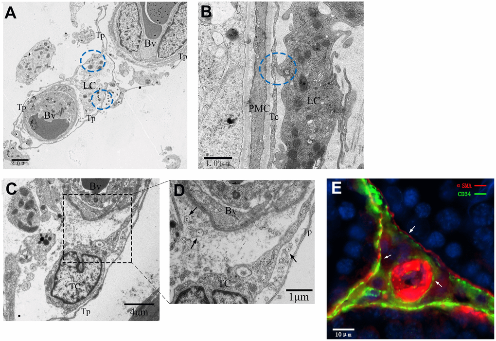
Figure 11.Interactive network of cells including telocytes in the inter-tubular space of rat testes. (A–D) Transmission electron micrographs show that telocytes form interactive networks by extending their telopodes to connect with Leydig cells and surrounding blood vessels (A, C) through various secretory vesicles (D, indicated by the arrows). The circles highlight the direct contact between telopodes and Leydig cells (A–B). (E) Immunofluorescence staining of CD34 (green) and αSMA (red) shows telocytes surrounding the PMC and the blood vessel. Arrows show the telocytes. Higher magnification illustrates the rectangular area. Tp: telopode; Bv: Blood vessel; LC: Leydig cells; PMC: peritubular myoid cells; TC: telocyte. Scale Bar = A: 2μm; B: 1μm; C: 5μm;D: 1μm; E: 10μm.