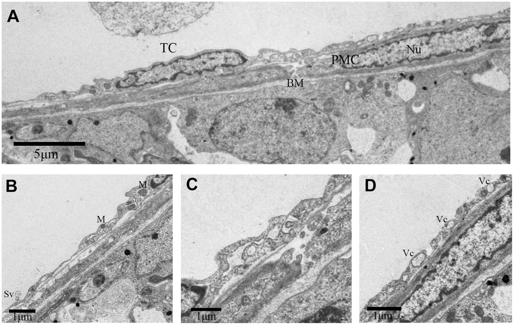
Figure 3.TEM photograph of a telocyte in rat testis. (A) Telocyte overlays the peritubular myoid cell in the lamina propria of seminiferous tubule. (B, D) Cytoplasmic process shows long extensions with (B) mitochondria and (D) vesicles. (C) The telopode seems more developed, with winding, thickened and dichotomous feature. TC: Telocyte; PMC: Peritubular myoid cell; Bm: Basement membrane; Nu: Nucleus; M: Mitochondria; Sv: Secretory vesicle; Vc: Vesicles. Scale Bar = A: 5μm, B–D:1μm.