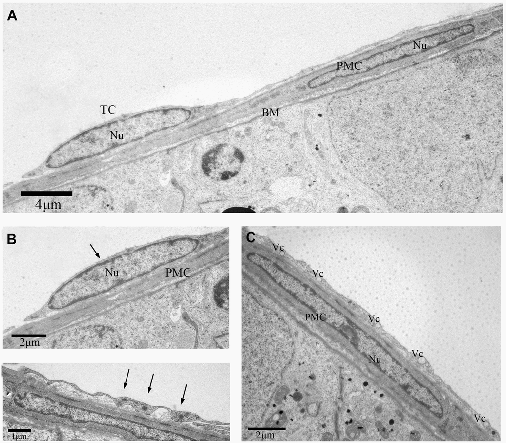
Figure 4.TEM photograph of another telocyte in the rat testis. (A) The telocyte surrounds the seminiferous tubule and the peritubular myoid cell.(B) The telocyte shows distinctly elongated nucleus (indicated by the arrow) with a small amount cytoplasm. (C) The cytoplasmic process shows few attachment plaques (indicated by the arrows). (D) The cytoplasmic prolongation shows many vesicles. TC: Telocyte; PMC: Peritubular myoid cell; Nu: Nucleus; Bm: Basement membrane; Vc: Vesicles. Scale Bar = A: 4μm; B:2μm; C: 1μm; D: 2μm.