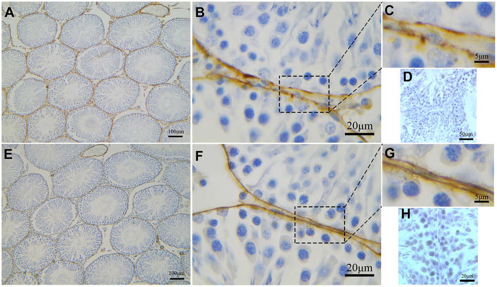
Figure 7.Immunohistochemical analyses of peritubular tissue in the rat testis with anti-αSMA and anti-CD34 antibodies. (A–C) CD34 positive staining (brown) surrounds the seminiferous tubules. Higher magnification (C) shows segmental pattern of CD34 staining. (E–G) αSMA positive staining (brown) also surrounds the seminiferous tubules. Higher magnification (G) shows smooth uniform staining pattern. Higher magnification illustrates the rectangular area. (D, H) shows negative contral group of rat testis. Scale Bar = A, E: 100μm; B, F: 20μm; C, G: 5μm; D: 50μm; H: 20μm.