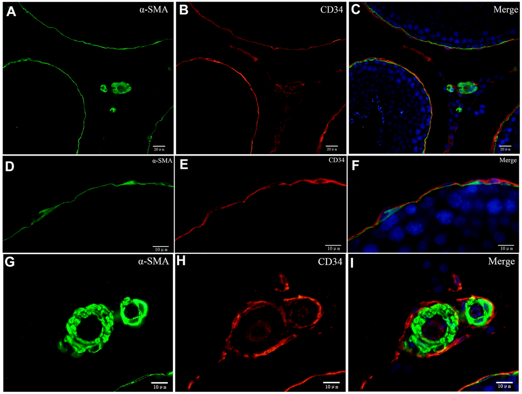
Figure 8.Double immunofluorescence of telocytes in the rat testis with anti-αSMA and anti-CD34 antibodies. (A–F) Immunofluorescence staining shows positive αSMA (A; green) and CD34 (B; red) staining of the inner and the outer layers surrounding the seminiferous tubules. (G–I) Immunofluorescence staining shows positive CD34 staining (red) around the blood vessel and in endothelial cells, and positive αSMA staining (green) on the vessel wall. Scale Bar = A–C: 20μm; D–I: 10μm.