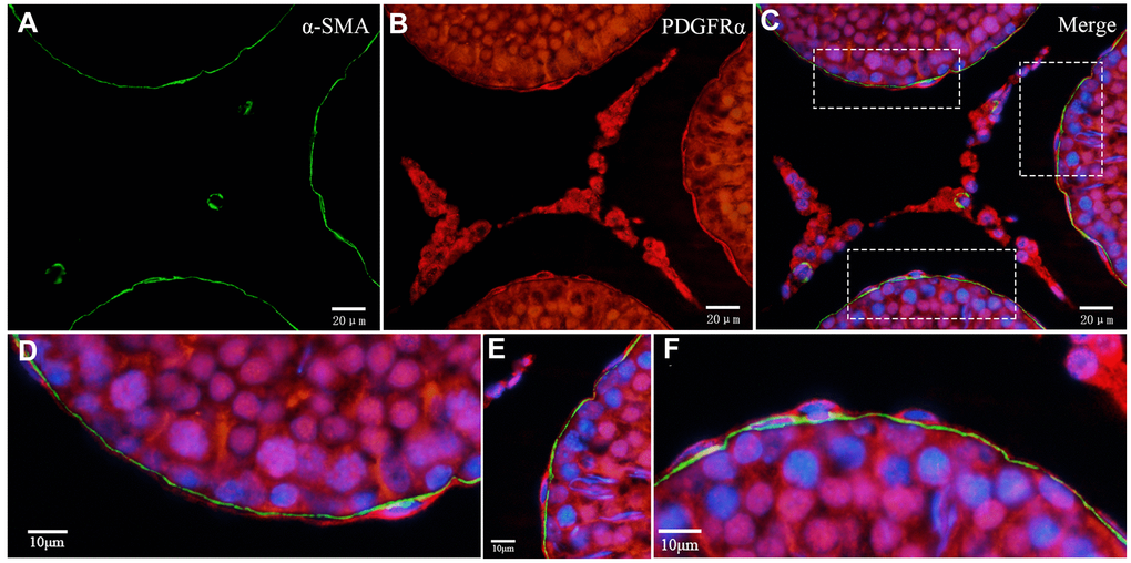
Figure 9.Double immunofluorescence staining of telocytes in the rat testis with anti-αSMA and anti-PDGFRα antibodies. (A–C) Immunofluorescence images show positive αSMA (green) and PDGFRα (red) staining inside/outside as continuous layers around the seminiferous tubule. (D–F) Immunofluorescence images show higher magnification illustrates the rectangular area in (C). Scale Bar = A–C: 20μm; D–F:10μm.