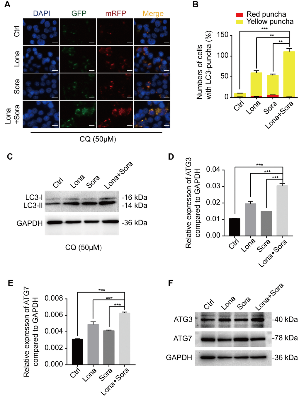
Figure 4.ATG3 is involved in the autophagic flux induced by lonafarnib and sorafenib co-treatment. (A) HepG2 cells were transfected with mRFP-GFP-LC3 reporter after treatment with lonafarnib (10 μM) and/or sorafenib (5 μM) plus CQ (50 μM) for 48 h. Microscopy images merged with GFP, RFP and DAPI fluorescence of representative cells. Scale bar = 10 μm. (B) The numbers of autophagosomes and autolysosomes were summarized and the data were presented as the means ± SD of triplicates. **P < 0.01; ***P < 0.001. (C) Western blot analysis of protein levels of LC3B. HepG2 cells were treated with lonafarnib and/or sorafenib in the presence of CQ (50 μM). (D) and (E) mRNA expression of ATG3 and ATG7 performed by qPCR assay. ***P < 0.001. (F) Western blot analysis of ATG3 and ATG7 protein. HepG2 cells were treated as indicated.