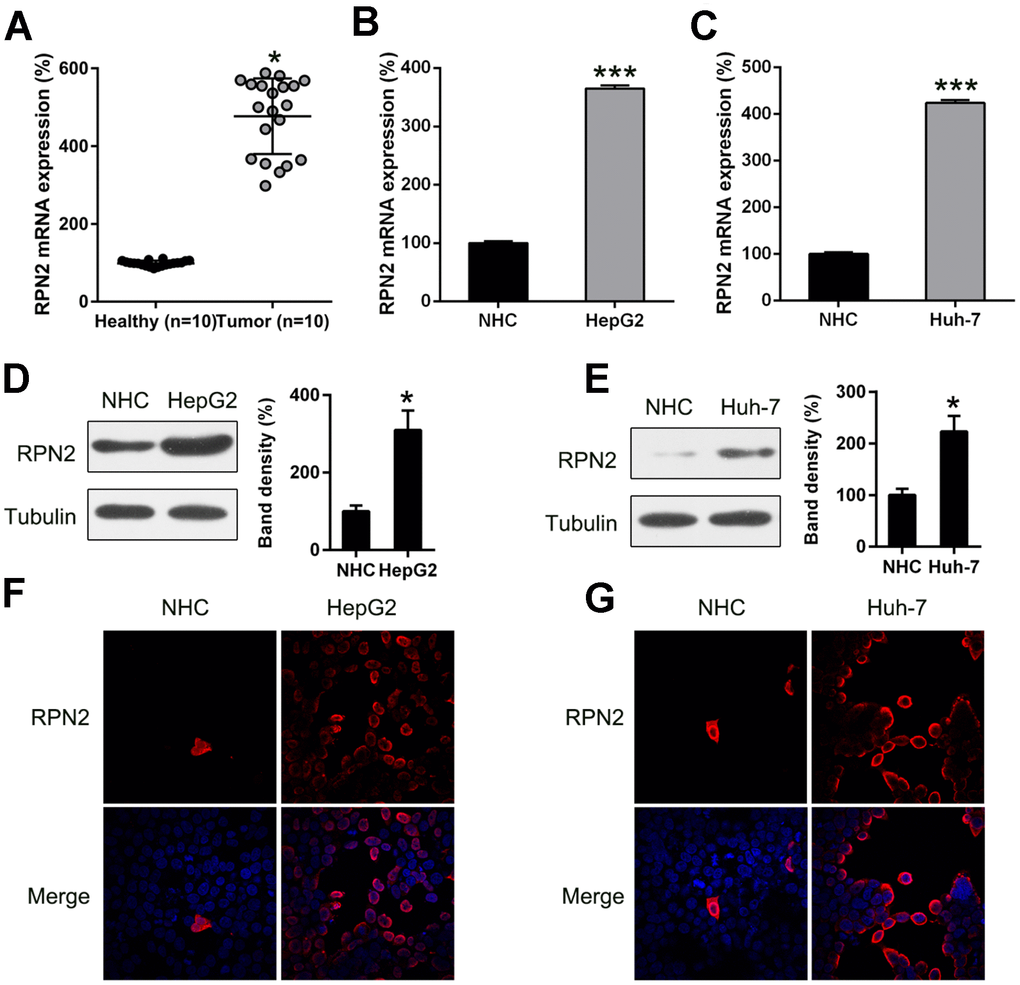
Figure 1.RPN2 expression in HCC cell lines and tissue specimens. (A) RPN2 expression in specimens obtained from HCC patients (n = 20) vs normal healthy tissue (n = 20). (B, C) mRNA level of RPN2 in Huh-7 and HepG2 cells was determined by qPCR. (D, E) Protein expression of RPN2 in HCC cell lines, Huh-7and HepG2, as well as normal hepatic cells (NHC) was detected by WB. (F, G) Subcellular localization of RPN2 in HCC cell lines, Huh-7 and HepG2, was detected by immunofluorescence. RPN2 and nuclear DNA were stained with anti-RPN2 antibody (red) and DAPI (blue) respectively. Merged image showed the subcellular localization of RPN2. Images were captured on a fluorescence microscope. The band of target protein was normalized to the density of action. The quantification was performed independently in a single band. The experiments were performed three times. Results are recorded as mean ± SD. *P < 0.05, ***P < 0.001 vs control group.