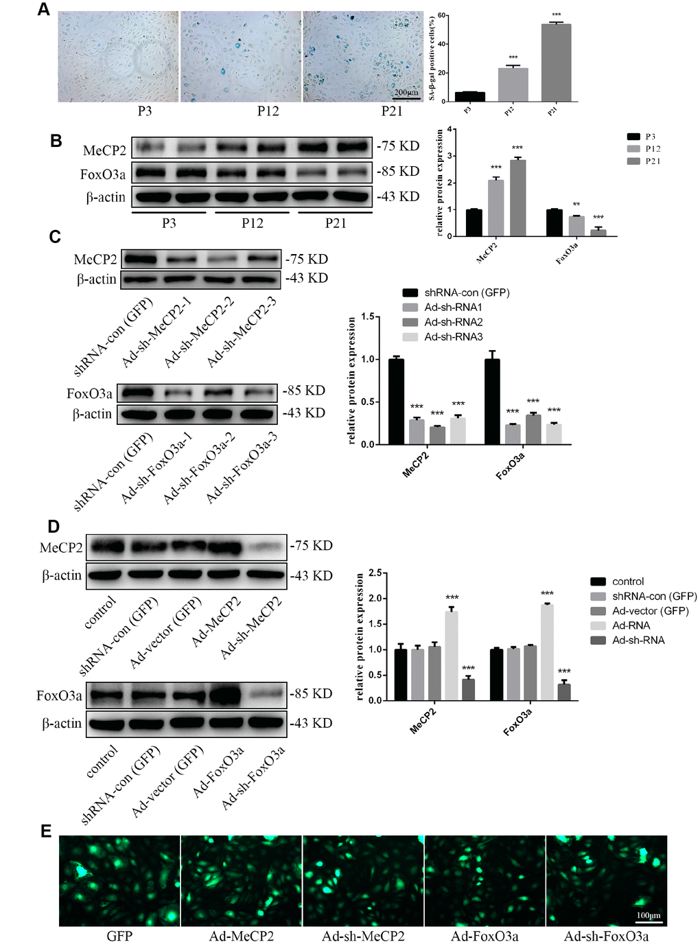
Figure 2.Protein expression during replicative aging and the efficacy of adenoviral vectors containing interference sequences. (A) Senescence-associated beta-galactosidase (SA-β-gal) staining during P3, P12, and P21 confirmed the increasing rate of aging EPCs during replicative senescence. (B) Protein levels of MeCP2 and FoxO3a were detected by western blotting during replicative senescence. (C) Protein levels of MeCP2 and FoxO3a were detected by western blotting after transfection with Ad-sh-MeCP2 or Ad-sh-FoxO3a. (D) Protein levels of MeCP2 and FoxO3a were detected by western blotting after overexpression and silencing of MeCP2 or FoxO3a. (E) Efficacy of adenoviral vectors was confirmed under fluorescence microscopy. *P < 0.05, **P < 0.01, and ***P < 0.001 vs. control.