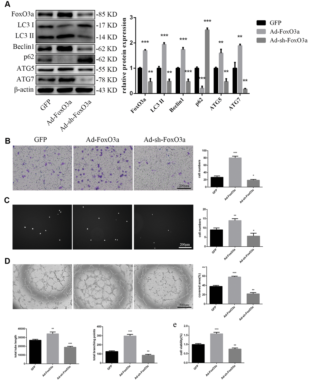
Figure 3.FoxO3a promoted autophagy and cellular function. (A) LC3 II, Beclin1, p62, ATG5, and ATG7 protein levels were detected by western blotting after transfection with Ad-FoxO3a or Ad-sh-FoxO3a for 48 h. (B) Cell migration was evaluated by Transwell migration assays after transfection with Ad-FoxO3a or Ad-sh-FoxO3a for 48 h. (C) Cell adhesion ability was evaluated by matrix adhesion assays after transfection with Ad-FoxO3a or Ad-sh-FoxO3a for 48 h. (D) Angiogenic ability was evaluated by Matrigel assays after transfection with Ad-FoxO3a or Ad-sh-FoxO3a for 48 h. (E) Cell viability was evaluated with CCK-8 after transfection with Ad-FoxO3a or Ad-sh-FoxO3a for 48 h. *P < 0.05, **P < 0.01, and ***P < 0.001 vs. control.