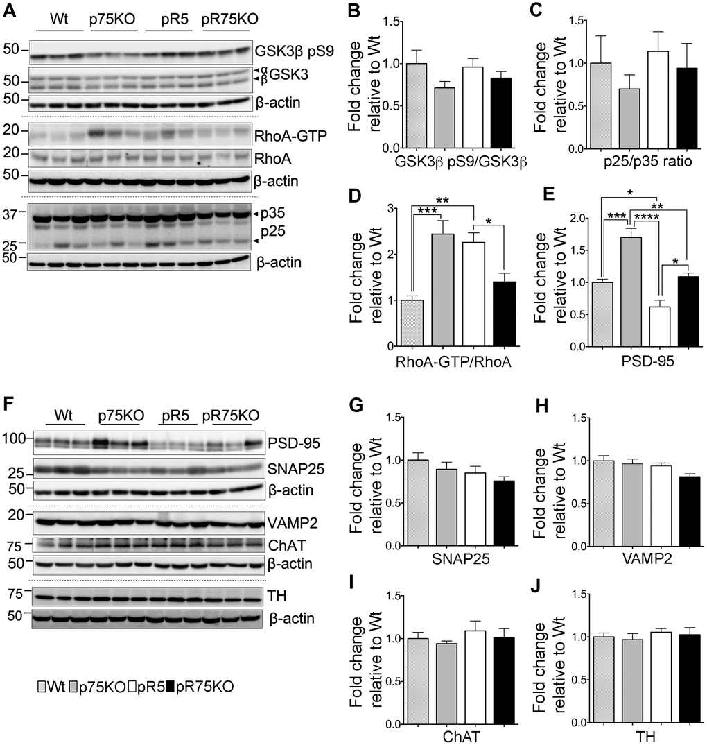
Figure 3.Synaptic proteins, neuronal markers and Tau kinase activity in pR75KO at 6 months. (A) Protein blots of kinases involved in Tau phosphorylation, GSK3β, RhoA and Cdk5 activators, p35 and p25 proteins in the forebrain of Wt, p75KO, pR5, and pR75KO mice. Protein band intensity quantification of inactive GSK3:GSK3β pS9 normalised with total GSK3β (B), Cdk5 activators, p25/p35 ratio (C), and active RhoA-GTP normalised with total RhoA (D). All band intensities showing B-D are expressed as fold change relative to Wt. F) Protein blots of post-synaptic protein, PSD-95 and pre-synaptic proteins, SNAP25 and VAMP2, tyrosine hydroxylase (TH) and choline acetyl transferase (ChAT). Protein band intensity quantification of PSD-95 (E), SNAP25 (G), VAMP2 (H), choline acetyl transferase (ChAT) (I), and tyrosine hydroxylase (TH) (J) normalised with total β-actin of respective blot and expressed as fold change relative to Wt. Data are represented as the mean ± SEM, n=6. Statistical comparisons were performed using one-way ANOVA and Tukey’s test. Statistical significance: *P<0.05, **P<0.01, ***P<0.001, ****P<0.0001.