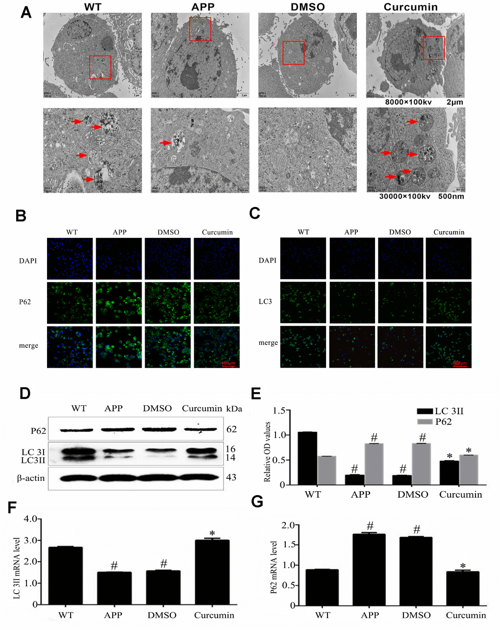
Figure 1.Curcumin induced autophagy in N2a/APP695swe cells. (A) Lesser and greater magnification transmission electron microscopy images for the whole cell body of each group. Arrows denote autophagic lysosomes and autophagosomes. (B) P62 and (C) LC3 immunofluorescence were used to measure the level of autophagy. Bar=100 μm. (D–E) Western blot analysis of LC3II, and p62 in each group. (F–G) Relative mRNA expression of (F) LC3 and (G) P62 of each group; The data represent as mean ± SEM of a typical series of 3 experiments (# P<0.05, compared to the WT group; * P<0.01, compared to the APP or DMSO group).