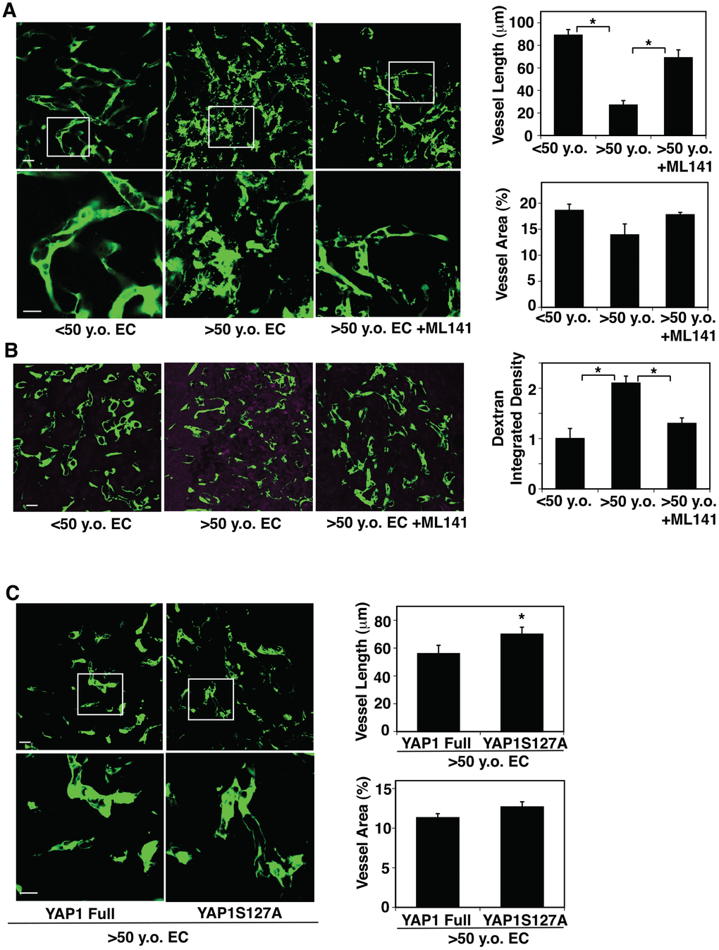
Figure 4.CDC42-YAP1 signaling mediates age-dependent decline in blood vessel formation in subcutaneously implanted gel. (A) IF micrographs showing vascular structures formed in the subcutaneously implanted fibrin gel supplemented with GFP-labeled ECs isolated from <50 y.o. or >50 y.o. human adipose tissues or in combination with treatment with ML141 (500 nM). Scale bar, 10 μm. Graphs showing quantification of vessel length (top) and vessel area (bottom) in the gel (n=7, mean ± s.e.m., *, p<0.05). (B) IF micrographs showing low MW fluorescently labeled dextran leakage (magenta) and GFP-labeled blood vessel formation (green) in the subcutaneously implanted fibrin gel supplemented with GFP-labeled ECs isolated from <50 y.o. or >50 y.o. human adipose tissues or in combination with treatment with ML141 (500 nM). Scale bar, 10 μm. Graph showing quantification of fluorescently labeled dextran leakage in the gel (n=7, mean ± s.e.m., *, p<0.05). (C) IF micrographs showing vascular structures formed in the subcutaneously implanted fibrin gel supplemented with GFP-labeled ECs isolated from >50 y.o. human adipose tissues in combination with treatment with retrovirus overexpressing full-length YAP1 or YAP1S127A mutant construct. Scale bar, 10 μm. Graphs showing quantification of vessel length (top) and vessel area (bottom) in the gel (n=7, mean ± s.e.m., *, p<0.05).