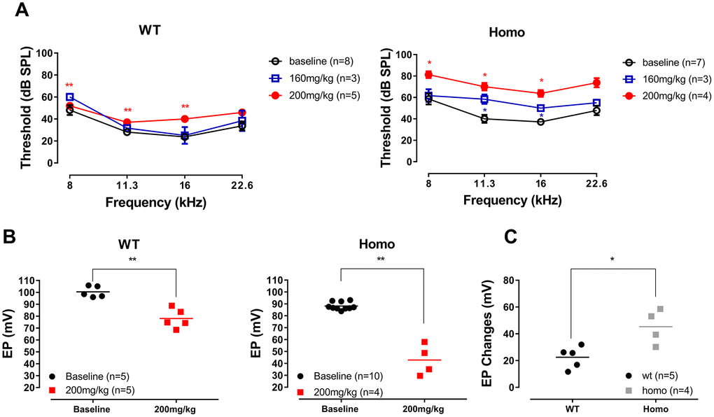
Figure 12.ABR and EP measurement before and after furosemide treatment. (A) ABR thresholds of four representative frequencies (8, 11.3, 16 and 22.6 kHz) monitored 20 to 30 minutes after furosemide injection. No threshold shift was observed in wild-type mice when 160 mg/kg furosemide was administered (all P>0.05, two-way ANOVA followed by Bonferroni post-test), and only a mild threshold elevation occurred when the dose was increased to 200 mg/kg (left panel, **all P<0.01 at 8, 11.3 and 16 kHz, two-way ANOVA followed by Bonferroni post-test). Homozygous mice responded to 160 mg/kg furosemide with moderate threshold elevation (right panel, *P=0.038, F(1,2)=25.00 at 11.3 kHz and P=0.020, F(1,2)=49.00 at 16 kHz, two-way ANOVA followed by Bonferroni post-test), and hearing loss developed when the dose increased to 200 mg/kg (right panel, *all P<0.05 at 8, 11.3, 16 kHz, two-way ANOVA followed by Bonferroni post-test). (B) EP measurements in the same animals 2 hours after the 200mg/kg furosemide injection (**P=0.0014, t=5.376 and df=6.403 for wild-type and P=0.0053, t=6.932 and df=3.149 for homozygous mice, Student’s unpaired t-test with Welch’s correction). Baseline EPs were collected from the age-matched mice from Figure 3. (C) EP changes in both groups compared to the averaged baselines of non-treated controls younger than 20 weeks old. Homozygous mice had a significantly larger EP reduction than wild-type mice (*P= 0.0287, t=3.084, df=4.825, Student’s unpaired t-test with Welch’s correction).
Figure 12 — Hearing consequences in Gjb2 knock-in mice: implications for human p.V37I mutation | Aging