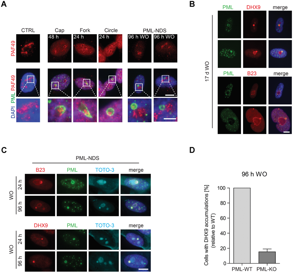
Figure 2.The association of specific subtypes of PNAs with different functional states of nucleoli. (A) The activity of RNAP I, evaluated as relocalization (segregation) of RNAP I subunit PAF49 and the presence of specific forms of PNAs were visualized by wide-field immunofluorescence microscopy of PAF49 (red) and PML (green) in RPE-1hTERT during different time-points of doxorubicin-treatment (0.75 μM) and its removal (WO). The insets show selected nucleoli with different states of RNAP I. Bars, 10 μm for whole cells and 4 μm for insets. (B) Long-term persistence (17 days after doxorubicin removal) of PML-NDS marked by PML (green) with accumulations of nucleolar proteins DHX9 and B23 (both in red). Bar, 10 μm. (C) Accumulation of B23 and DHX9 inside PML-NDS was visualized by immunostaining with respective antibodies (accumulated proteins – red, PML – green). Nuclei (A–C) and nucleoli (C) were visualized by DAPI (blue) and TOTO-3 (cyan), respectively. The images were captured with 63×/1.4 objective. Bar, 10 μm. (D) RPE-1hTERT PML-WT and PML-KO cells were treated with 0.75 μM doxorubicin for 48 hours; after that doxorubicin was removed and the cells were further cultured. 96 hours after doxorubicin washout the cells were fixed, stained with antibodies against DHX9 and PML and imaged with the Olympus ScanR microscope. The occurrence of cells with DHX9 accumulations were analyzed by the ScanR analysis software. The relative occurrence of cells with accumulations of DHX9 is shown. Two biological replicates were evaluated. Results are presented as a mean ± s.d.