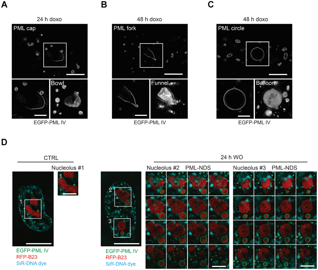
Figure 3.Three-dimensional reconstruction of structural subtypes of PNAs. High resolution live-cell structured illumination microscopy of the cap- (A), fork- (B) and circle-like (C) PNAs of RPE-1hTERT cells stably expressing the EGFP-PML IV isoform harvested 24 and 48 hours after doxorubicin-treatment. Central layer of the whole cell (upper images; bar, 5 μm) and central layer of respective type of PNAs (lower-left images; bar, 2 μm) are shown together with reconstructed 3-D images (lower-right images) from 28 (cap), 30 (fork) and 54 (circle) layers using ImageJ 3D viewer plugin. (D) High resolution live-cell SIM images of RPE-1hTERT stably expressing EGFP-PML IV and RFP-B23. A control untreated cell (left) and a cell containing PML-NDS, imaged 24 hours after doxorubicin washout (right). The central layer of whole cells (bar, 10 μm) are shown together with three insets of nucleoli (1 layer for the nucleolus of control cell and 16 layers for the nucleoli with adjacent PML-NDS; bar, 4 μm). DNA was labeled with SiR-DNA dye.