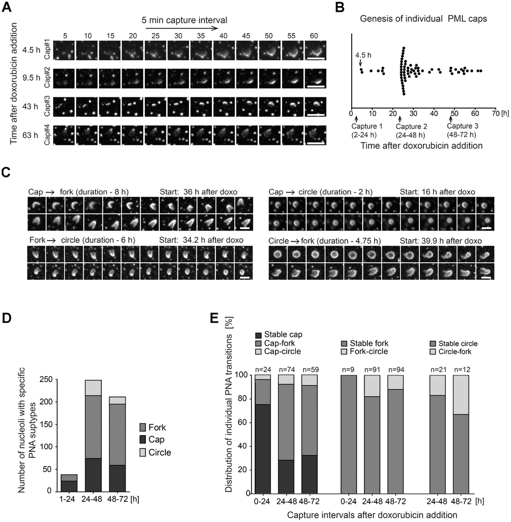
Figure 4.Structural transitions of PNAs subtypes. RPE-1hTERT stably expressing EGFP-PML IV were analyzed by live-cell imaging in three consecutive capturing sessions spanning 2–24, 24–48 and 48–72 hours after doxorubicin treatment (0.75 μM). (A) Series of sequential images of four individual cap formations (cap#1–4) that initiated 4.5, 9.5, 43 and 63 hours after doxorubicin addition. (B) Plot of all genesis of PML-caps recorded during three capturing intervals. Dots represent de novo formation of individual PML-caps. (C) Time-dependent evolution and transition of subtypes of PNAs. The four characteristic transitions – 'cap-to-fork', 'cap-to-circle', 'fork-to-circle' and 'circle-to-fork' – are represented by series of sequential images. The initiation of capturing and the length of recorded time (in parentheses) are given for each type of transition. (D) The quantitative distribution of specific subtypes of PNAs in three capturing sessions (see Figure 1B for comparison). (E) The quantitative distribution of specific PNAs subtype transitions in three capturing sessions. The PNAs that did not change for over 5 hours were considered as stable. Bars, 4 μm.