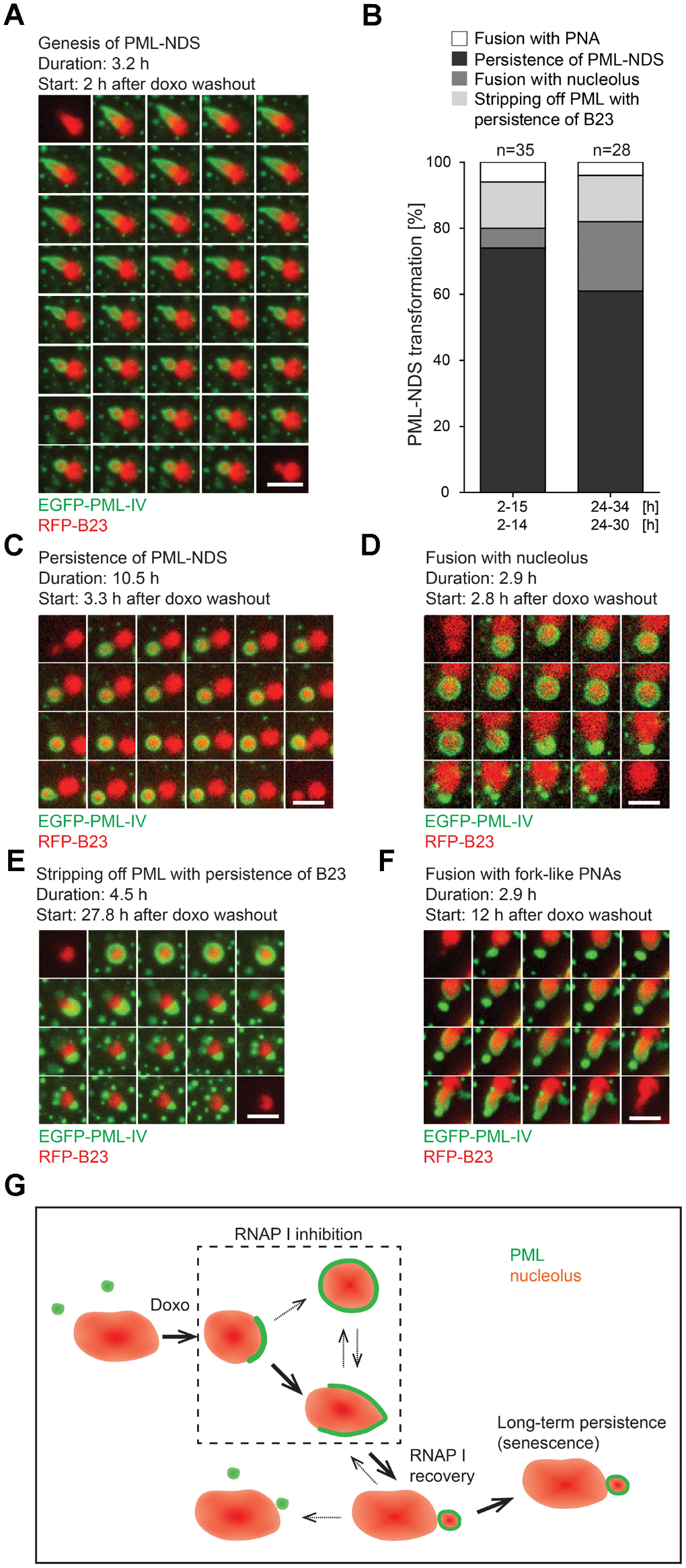
Figure 5.The genesis, stability and fate of PML-NDS. RPE-1hTERT stably expressing EGFP-PML IV (green) and RFP-B23 (red) were treated with 0.75 μM doxorubicin for 48 hours and analyzed by live-cell imaging after doxorubicin washout (up to 34 hours) for the presence of PML-NDS. (A) Series of sequential images mapping the genesis of PML-NDS. (B) Comparison of proportional representation of PML-NDS fates between two time-lapse capturing sessions (experiment I: 2–15 and 24–34 hours; experiment II: 2–14 and 24–30 hours) after drug removal. Four different fates of PML-NDS were monitored: persistence (no change) (C), fusion with nucleolus (D), stripping of PML with B23 persistence (E), and fusion with fork-like PNAs (F). EGFP-PML IV, green; RFP-B23, red; bars, 4 μm. The initiation of capturing and the length of recorded time are given for each type of transition. (G) Schematic representation of PNAs transmutations recorded by time-lapse microscopy. The bold arrows show the main transition pathways observed.