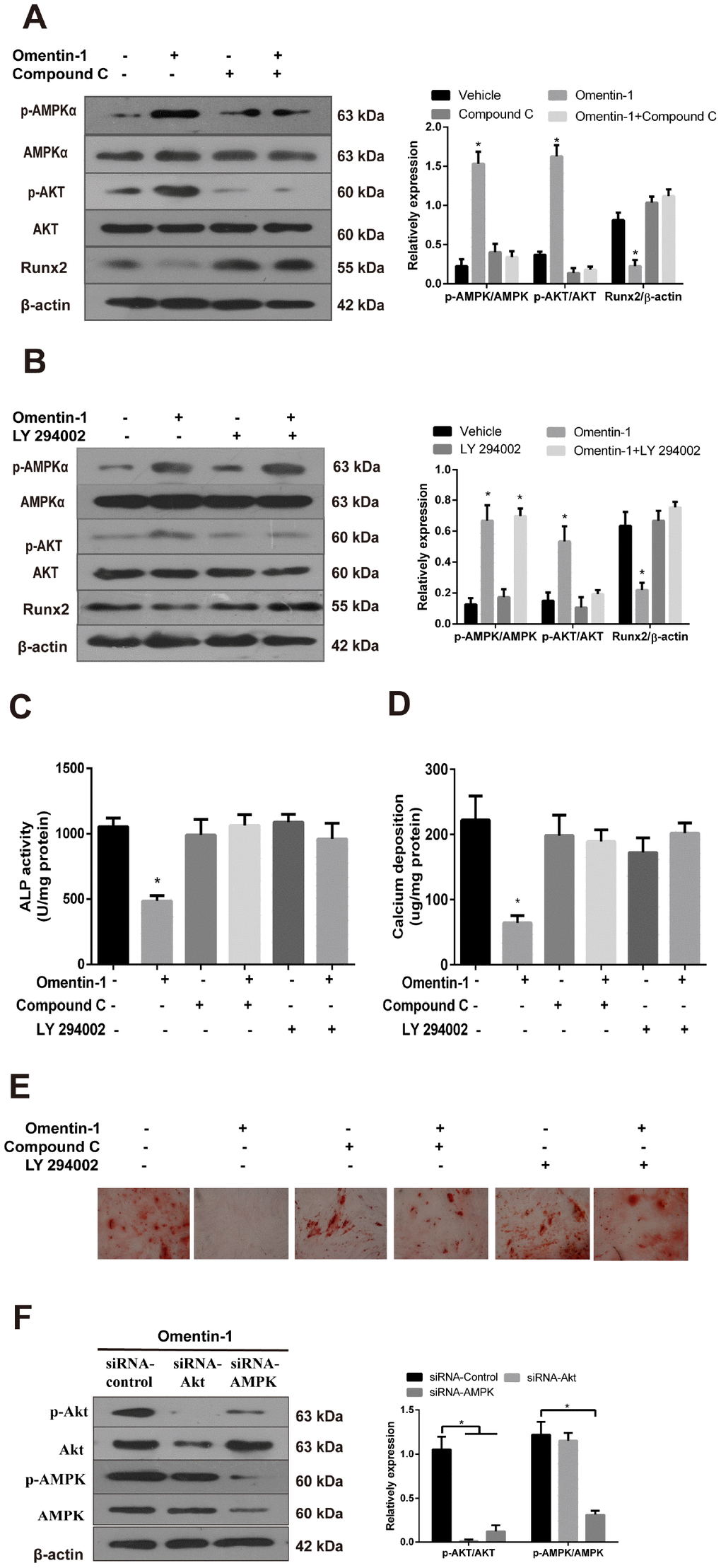
Figure 4.The AMPK/Akt signaling pathway mediates omentin-1-induced inhibition effects on osteoblastic differentiation and mineralisation of CVSMCs. CVSMCs were stimulated by Compound C (10 μM) or LY 294002 (30 μM) for 30 min and then treated with omentin-1 (400 ng/ml) for 48 hours. The cell lysates were tested by western blot and incubated with antibodies against p-AMPKα, AMPKα, p-Akt, Akt and Runx2. (A, B) Representative results were shown in the left panel and the data were presented as densitometric ratios of p-AMPK/AMPK, p-Akt/Akt and Runx2/β–actin respectively (lower panel). (C) The ALP activity was measured by ALP kit. Results are represented by mean ± SD with 3 replicates for each group. (*p < 0.05). One-way ANOVA with the Tukey’s HSD post hoc analysis was adopted. (D, E) CVSMCs were treated with Compound C (10 μM) or LY 294002 (30 μM) for 30 min and then treated with omentin-1 (400 ng/ml) every two days for a period of 14 days. Calcium deposition was tested (D) and represented microscopic pictures of Alizarin Red S staining view were shown (E). (F) Expression of p-AMPK and p-Akt in CVSMCs after AMPK or Akt knockdown were analyzed by western blot. All Results are represented by mean ± SD with 3 replicates for each group. One-way ANOVA with the Tukey’s HSD post hoc analysis was adopted.