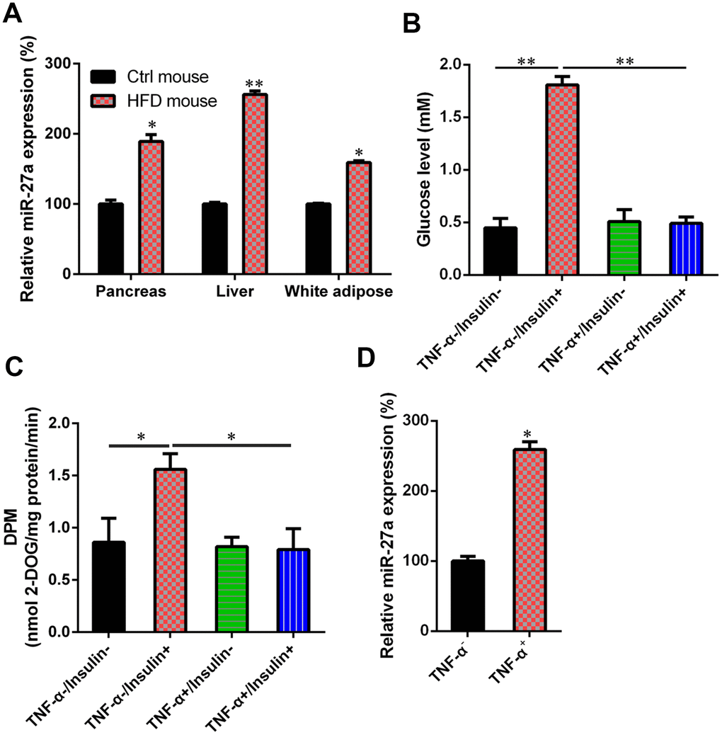
Figure 1.Upregulation of MiR-27a in HFD-fed mice and IR cells. (A) For obese mice fed with HFD, an increase in miR-27a expression levels in pancreas, liver, and white adipose tissue was revealed using real-time PCR at week 11 after modeling. (B) Cells were treated with high-glucose DMEM containing FBS (10%, w/v) supplemented with TNF-α (10 ng/ml) for one day. Subsequently, they were incubated for 0.5 h with another cell medium, i.e., insulin (100 nM) in high-glucose DMEM containing FBS (10%, w/v). The establishment of the IR adipocyte model was confirmed by glucose level results. (C) 2-deoxyglucose uptake assay was also performed to detect the glucose level in both cell and cell culture medium. (D) qPCR was utilized to showed that the miR-27a levels were obviously increased in the IR cell model treated with TNF-α, compared with those of the normal 3T3-L1 cells. Expression data from each mouse was normalized to that of randomly assigned mouse in control group. Number of animal per group = 8. **P < 0.01, *P < 0.05, compared to indicated groups.