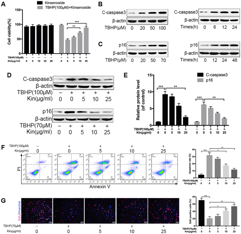
Figure 1.Kin suppresses NPC apoptosis and senescence under oxidative stress. (A) The NPCs were treated with different concentrations of Kin (0, 5, 10, 25 or 50 μg/ml) alone for 24 h, or Kin (0, 5, 10 or 25 μg/ml) for 2 h before receiving TBHP (100 μM) for 24 h. The cell viability was assessed by CCK-8. (B) The western blotting of C-caspase3 in the NPCs treated with different concentrations of TBHP (0, 20, 50 or 100 μM) for 24 h or TBHP (100 μM) for indicated time points (0, 6, 12 or 24 h). (C) The western blotting of p16 in the NPCs treated with different concentrations of TBHP (0, 20, 50 or 70 μM) for 48 h or TBHP (70 μM) for indicated time points (0, 12, 24 or 48 h). (D, E) The western blotting and quantitative protein levels of C-caspase3 and p16 in the NPCs as treated above. (F) The apoptosis rate in NPCs, treated with different concentrations of Kin (0, 5, 10 or 25 μg/ml) for 2 h before receiving TBHP (100 μM) for 24h, was analyzed by flow cytometry using Annexin V-APC/PI. (G) The cell proliferation was determined by quantification of EdU-positive cells in the NPCs treated with different concentrations of Kin (0, 5, 10 or 25 μg/ml) for 2 h before receiving TBHP (70 μM) for 24 h; scale bar: 100 μm. All data are expressed as mean ± SD of at least three independent experiments.