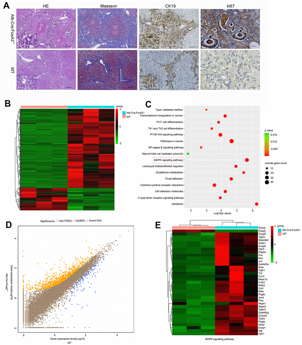
Figure 3.Loss of FoxA2 promotes the development of intrahepatic bile duct neoplasms and enhances MAPK-related gene expression. (A) Hematoxylin and eosin staining showed remarkable bile duct neoplasm formation in FoxA2-/- mice. Significant liver cirrhosis and proliferation (Ki67 immunohistochemistry) were also observed in the bile duct in FoxA2-/- mice (n=3 each group); (B) Heatmap showing the most differentially expressed genes (DEGs) between FoxA2-/- mice and WT mice. (n = 3 samples per group); (C) KEGG analysis of biological processes. Most of these DEGs were clustered in the “MAPK signaling pathway” category, followed by the “Pathways in cancer”, and “PI13K-AKT signaling pathway”, and so on. The bar indicates the P value; the threshold of P = 0.015 is shown; (D) Volcano plot of P values as a function of the weighted fold change for mRNAs; 805 genes were upregulated genes and 154 were down regulated in FoxA2-/- mice compared with WT mice.; (E) the activation of MAPK-signaling-related genes expression in FoxA2-/- mice.