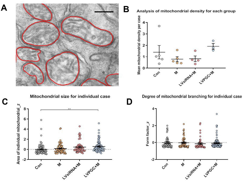
Figure 5.Electron microscopy observation of the destruction of mitochondrial structure in SN neurons. (A) Using electron microscopy (EM), SN images were collected from the group of con (number of mice, n=5), M (number of mice, n=5), LVPGC+M (number of mice, n=3), and LVsiRNA+M (number of mice, n=3). A total of 281 mitochondria were manually outlined and analyzed using QuPath software. Scale bar, 300 nm. (B) The average level of mitochondrial density was calculated for individual mice, which showed a higher trend in mitochondrial density in the LVPGC+M group and a lower trend in the M and LVsiRNA+M group compared to that in controls (P>0.05). (C) Mitochondrial size in the LVPGC+M group was significantly increased compared with that in control mice (P>0.01). (D) The degree of mitochondrial branching was also compared via the calculation of individual mitochondrial form factors.