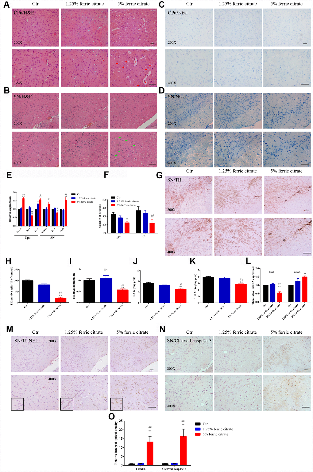
Figure 3.Iron overload induced by ferric citrate supplementation causes neurotoxicity in the SN and CPu. (A and B) Representative images of H&E staining display the histopathological damage in the CPu and SN induced by ferric citrate supplementation. Red arrows show white matter edema, red stars show vasodilatation, and green arrows display nerve cell swelling. (C, D and F) Representative images and quantification of NISSL staining display the decreased numbers of neurons in ferric citrate-supplemented mice. Error bars indicate SD. (E) qRT-PCR showed increased mRNA levels of TNF-α and IL-6 and decreased expression of IL-4 in the Cpu and SN of mice supplemented with ferric citrate (N=5). Error bars indicate SEM. (G and H) Representative images and quantification of TH staining display decreased numbers of dopaminergic neurons in the SN of mice supplemented with ferric citrate. Error bars indicate SD. (I) qRT-PCR shows decreased mRNA levels of TH in the SN of mice supplemented with ferric citrate (N=5). Error bars indicate SEM. (J and K) Quantifications show the decreased levels of DA and DOPAC in mice supplemented with ferric citrate. Error bars indicate SEM. (L) qRT-PCR show the mRNA levels of DAT and a-syn in the SN of mice supplemented with ferric citrate (N=5). Error bars indicate SEM. (M to O) Representative images and quantification from TUNEL and cleaved caspase-3 staining display the increased neuronal apoptosis in the SN of mice supplemented with ferric citrate. Bars, 100 μm. Compared with the Ctr group, *p<0.05 and **p<0.01. Compared with the 1.25% ferric citrate group, #p<0.05 and ##p<0.01.