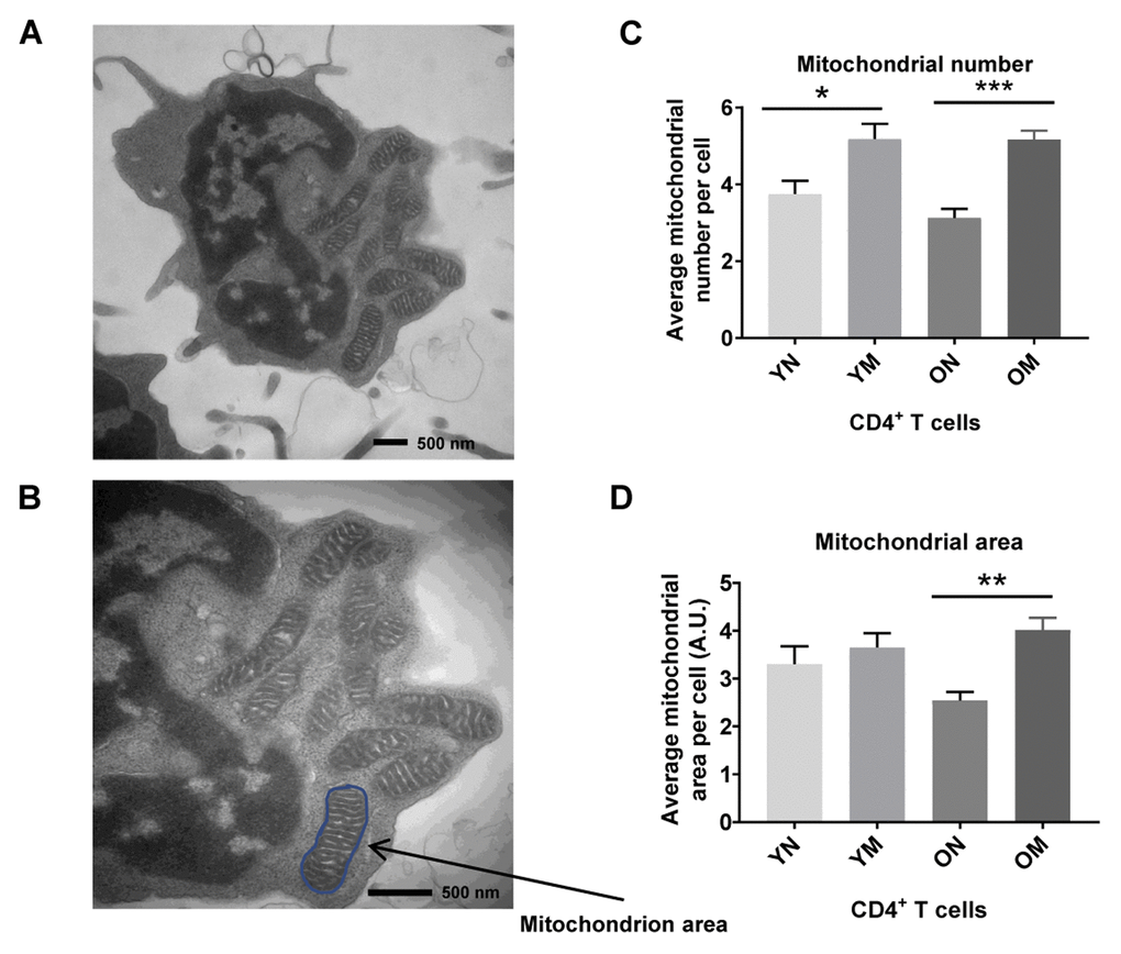
Figure 4.(A) TEM image of CD4+ T cell showing mitochondria, and (B) mitochondrion area (size) outlined in blue. (C) Memory (M) CD4+ T cells had a significantly higher mitochondria number than naïve (N) cells in cells from both young (Y) and old (O) (*p = 0.0267, ***p = 0.0008, respectively). (D) In memory CD4+ T cells of both young and old, the mitochondrial area was significantly greater compared to naïve CD4+ T cells (**p = 0.0030). Arbitrary Unit (A.U.). (C, D) P-values were calculated by Student’s t-test (two-tailed) using GraphPad PRISM 7 software. Error bars reflect the standard error of the mean (±SEM). N = 5 young, 4 old donors.