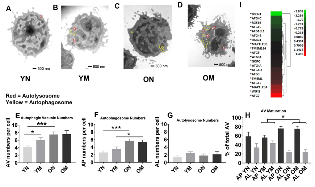
Figure 5.(A–D) Representative images showing autophagosomes and autolysosomes from young (Y) and old (O) naïve (N) and memory (M) CD4+ T cells. Circles in red indicate autolysosomes, in yellow indicate autophagosomes. (E) The number of autophagic vacuoles, the combination of autophagosomes and autolysosomes, was increased in naïve CD4+ T cells from older as compared to younger individuals (***p = 0.0006) and also increased in memory CD4+ T cells from young compared to naïve CD4+ T cells from young individuals (*p = 0.0424). (F) Autophagosomes were significantly higher in both naïve and memory CD4+ T cells from older individuals compared to naïve and memory CD4+ T cells of younger individuals (***p = 0.0001, *p = 0.0217, respectively). (G) No significant differences were found in the number of autolysosomes from young and old naïve and memory CD4+ T cells. (H) Significant differences were found in autophagic vacuole (AV) maturation of autophagosomes (AP) transitioning to autolysosomes (AL) between memory CD4+ T cells from older individuals compared to younger individuals (*p = 0.022). No differences were found for autophagic vacuole maturation for naïve CD4+ T cells. (E–H) P-values were calculated by Student’s t-test (two-tailed) using GraphPad PRISM 7 software. Error bars reflect the standard error of the mean (±SEM). N=5 old, 4 young donors. (I) Gene expression data for autophagy pathway genes in CD4+ T cells from older compared to young individuals showing up-regulation (above 0-1.5 Z- ratio) of some autophagy-related genes (ATG7, WIPI1, MAP1LC3A, ATG12, TMEM1, ATG3, ATG4D). Interestingly, BECN1 was down-regulated (-2.8 Z-ratio) in older compared to young individuals. N= 8 young, 25 old donors.