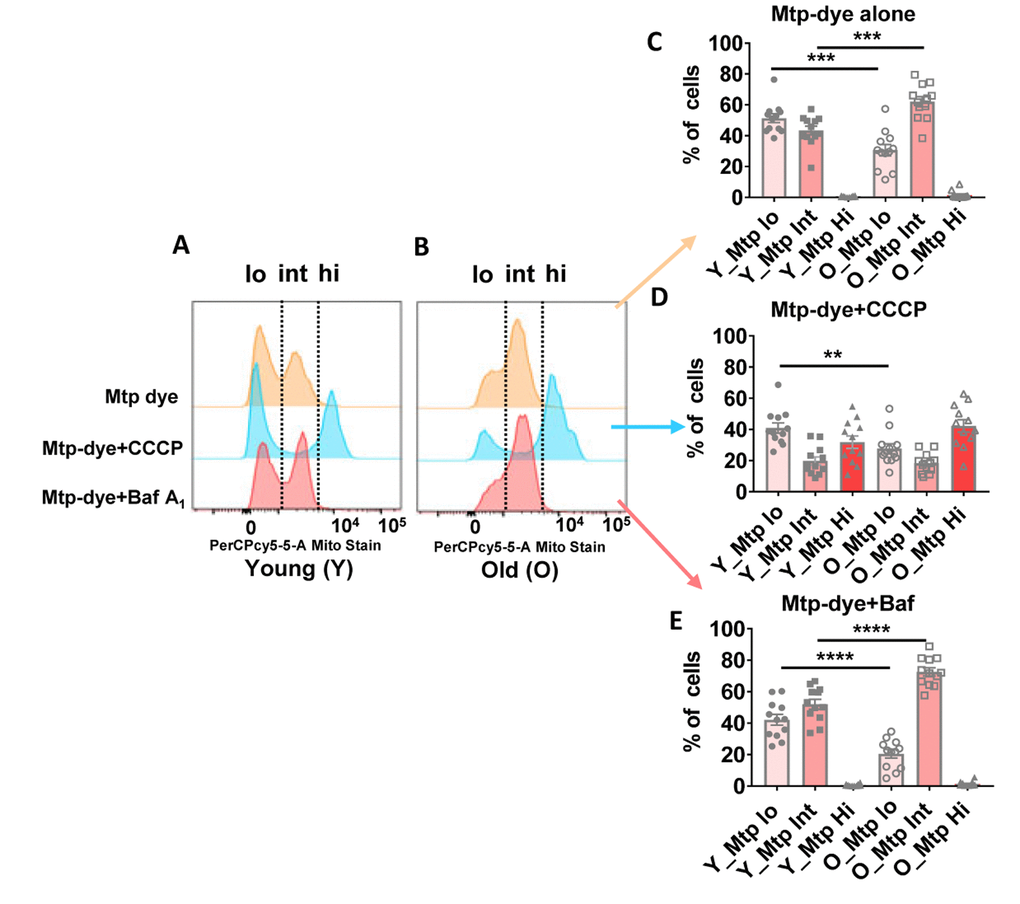
Figure 6.(A and B) Representative histograms from a young and old donor for all conditions using Mtphagy Dye (20,000 cells recorded). Summary data showing percentage of CD4+ T cells of young and old donors divided by fluorescent intensity (low [‘lo’], intermediate (‘int’), and high [‘hi’]) of Mtphagy Dye using, © Mtphagy Dye alone, (D) CCCP administration, and (E) Bafilomycin A1 administration. ****p = 0.0001, ***p = 0.0002 and 0.0003, **p = 0.0081. (C–E) P-values were calculated by Student’s t-test (two-tailed) using GraphPad PRISM 7 software. Error bars reflect the standard error of the mean (±SEM). N = 12 young, 12 old donors.