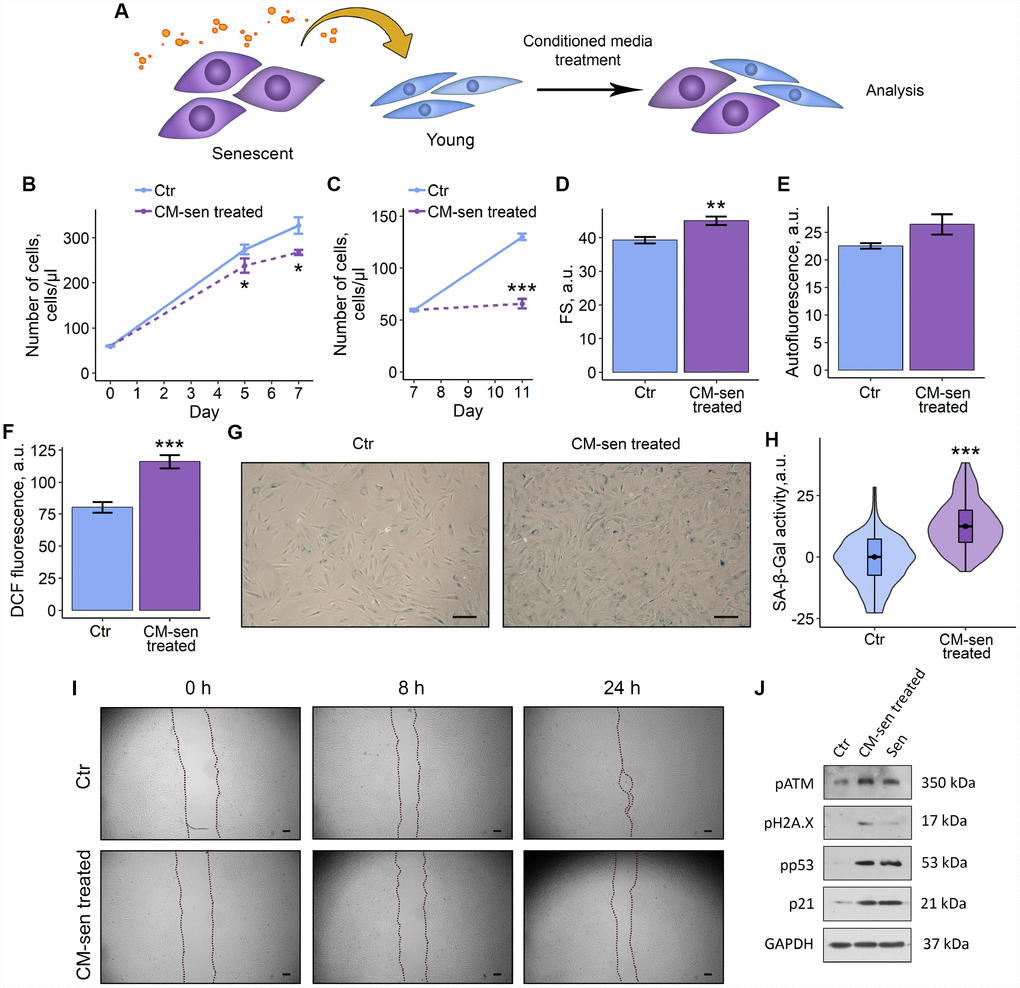
Figure 3.SASP from senescent ESCs triggers senescence in young cells. Ctr – young ESCs cultured in standard conditions. CM-sen treated – ESCs exposed to condition medium from senescent cells. Sen – senescent ESCs. (A) Experimental scheme of ESCs CM-sen treatment. (B) and (C) Growth curves of ESCs before and after reseeding, respectively. Cell number was determined by FACS at the indicated time points. (D–F) Cell size, autofluorescence and intracellular ROS levels of ESCs determined by FACS after 9 d of CM-sen treatment. Forward scatter (FS) reflects the average cell size, DCF fluorescence reflects ROS levels by oxidation of H2DCF-DA. Values are M ± S.D. (N=3). * – p<0.05, ** – p<0.01, *** – p<0.005 by Student’s t-test. (G) SA-β-Gal staining of Ctr and CM-sen treated ESCs. After 7 d of treatment ESCs were reseeded and additionally cultured for 3 d in order to perform staining of non-confluent cultures. (H) Quantification of SA-β-Gal activity values (G). Values presented as M and 95 % C.I. (N=100). *** – p<0.005 by Mann-Whitney test. (I) Wound healing analysis of ESCs cultured in standard conditions or pre-exposed to CM-sen for 4 d. Cells’ monolayers were scratched and migration activity of cells were estimated at the indicated time points. Scale bars of all images are 500 μm. (J) Western blot analysis of ATM, H2A.X and p53 phosphorylation levels and p21 protein expression performed after 7 d of treatment. Representative results of the three experiments are shown in the Figure. GAPDH was used as loading control.